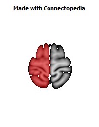

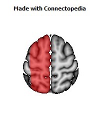
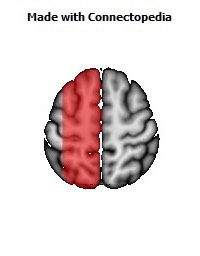
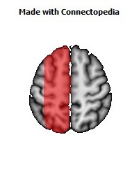
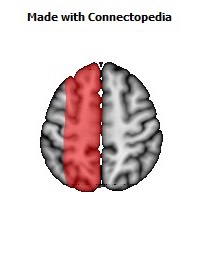
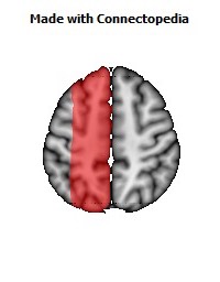
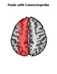
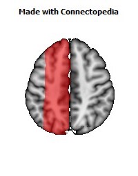
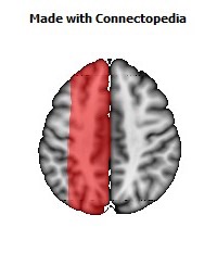
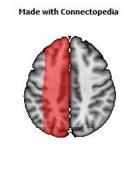
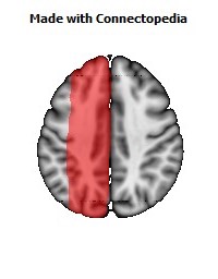
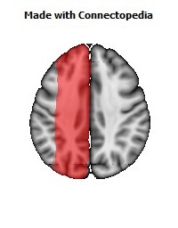
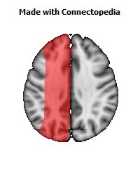
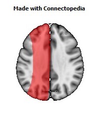
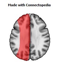
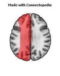
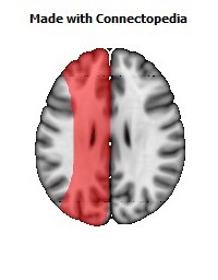
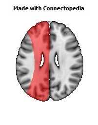
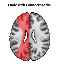
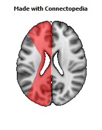
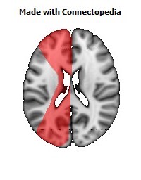
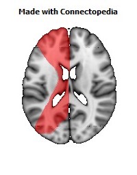
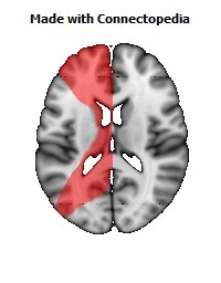
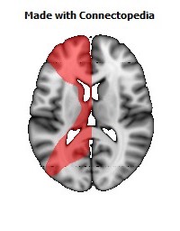
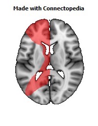
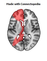
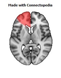
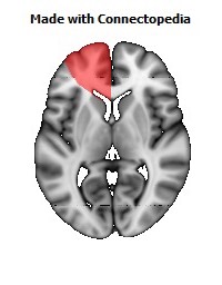
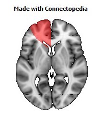
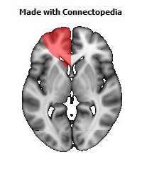
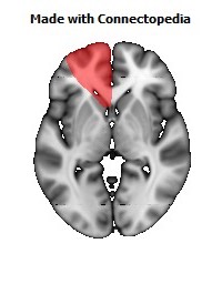
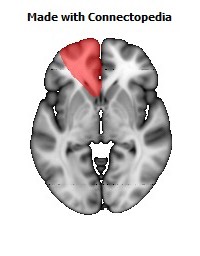
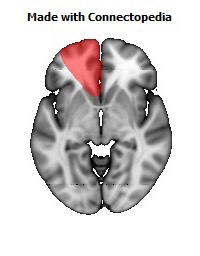

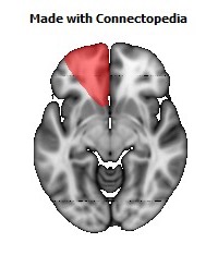
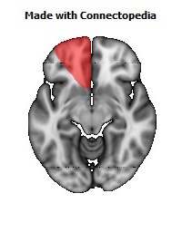
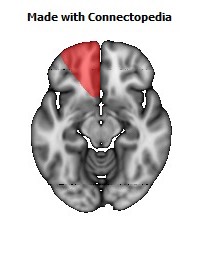
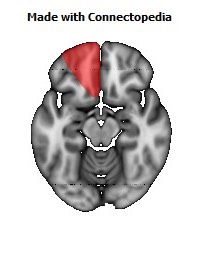
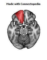
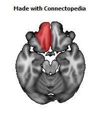
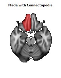
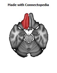


The anterior cerebral artery (ACA) is one of a pair of arteries on the brain that supplies oxygenated blood to most medial portions of the frontal lobes and superior medial parietal lobes. The two anterior cerebral arteries arise from the internal carotid artery and are part of the Circle of Willis.
The left and right anterior cerebral arteries are connected by the anterior communicating artery.
Structure
The ACA is classified into 5 segments with the smaller branches from the ACA "callosal" arteries (supracallosal) considered as the A4 and A5 segments:
- A1: this segment originates from the internal carotid artery and extends to the anterior communicating artery (AComm). The anteromedial central (medial lenticulostriate) arteries arise from this segment as well as the AComm, which irrigate the caudate nucleus and the anterior limb of the internal capsule
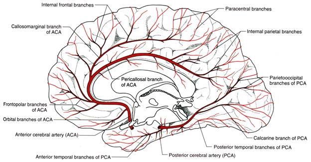
- A2: this segment extends from the AComm to the bifurcation forming the pericallosal and callosomarginal arteries. The recurrent artery of Heubner (distal medial striate artery), which irrigate the internal capsule, usually arises at the beginning of this segment near the AComm. 4 branches arise from this segment, the:
- Orbitofrontal artery (medial frontal basal): Arises first a small distance away from the AComm
- Frontopolar artery (polar frontal): Arises after the orbitofrontal close to when A2 curves posteriorly over the corpus callosum. Could also originate from the callosal marginal.
- A3: Also termed the pericallosal artery this is one of (or the only) the main terminal branches of the ACA, which extends posteriorly in the pericallosal sulcus to form the internal parietal arteries (superior, inferior) and the precuneal artery. This artery may form an anastomosis with the posterior cerebral artery.
- Callosal marginal artery: A commonly present terminal branch of the ACA, which bifurcates from the pericallosal artery. This artery in turn branches into the medial frontal arteries (Anterior, Intermediate, Posterior), and the paracentral artery, with the cingulate branches arising throughout its length. Depending on anatomical variation, the callosal marginal artery may be none discrete or not be visible. In the latter case, the branches mentioned will originate from the pericallosal artery. In a study of 76 hemispheres, the artery was present in only 60% of the cases. Angiography studies cite that the vessel can be seen 67% or 50% of the time.
Function
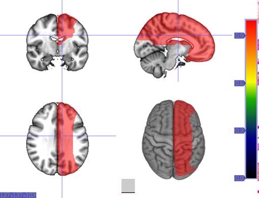
Areas supplied by the anterior cerebral artery include:
- The medial surface of the frontal lobe by the medial orbito-frontal artery, and parietal lobes
- The anterior four- fifths of the corpus callosum
- Approximately 1 inch of the lateral surfaces of frontal and parietal lobes, next to the medial longitudinal fissure
- Anterior portions of the basal ganglia and internal capsule
- Olfactory bulb and tract
Clinical relevance
Occlusion
Occlusion of the anterior cerebral artery may result in the following defects. If stroke occurs prior to the anterior communicating artery it is usually well tolerated secondary to collateral circulation. Occlusion of A2 segment(Post communal segment of Anterior cerebral artery) the following signs and symptoms may be noted.
- Paralysis or weakness of the contralateral foot and leg due to involvement of Motor leg area
- Cortical Sensory loss in the contralateral foot and leg
- Gait apraxia Impairment of gait and stance
- Abulia akinetic mutism, slowness and lack of spontaneity
- Urinary incontinence which usually occurs with bilateral damage in the acute phase
- Frontal Cortical release reflexes: Contralateral grasp reflex, sucking reflex, gegenhalten(paratonic rigidity)