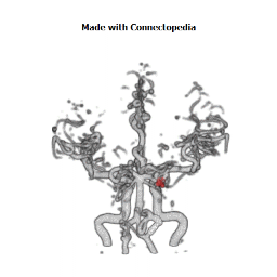

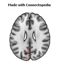

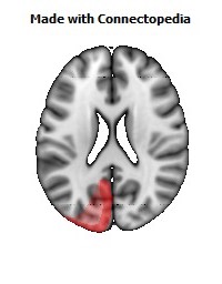
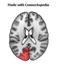
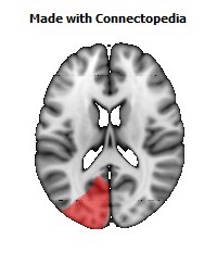
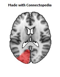
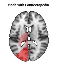
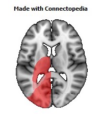
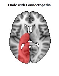
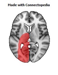
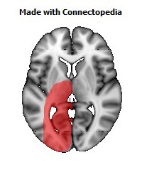
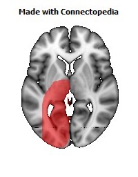
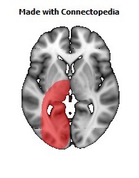
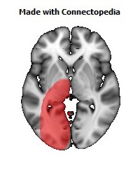
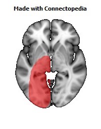
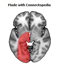
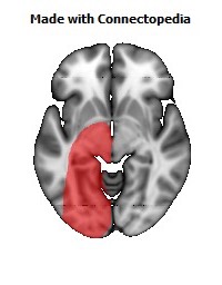
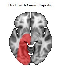
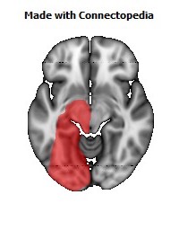
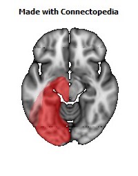
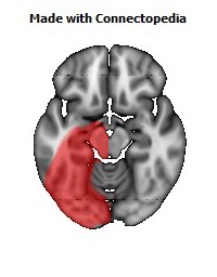
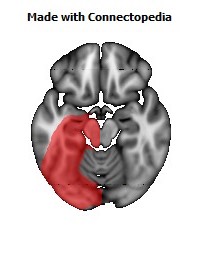
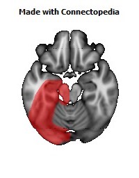
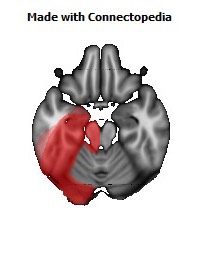
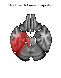
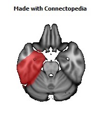
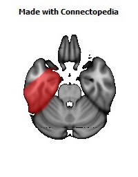

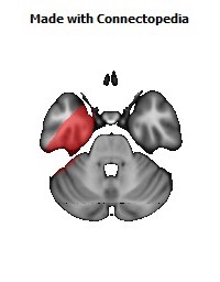
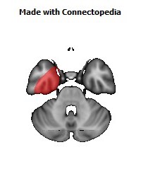
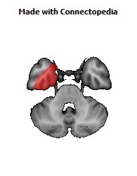
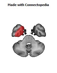
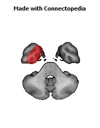
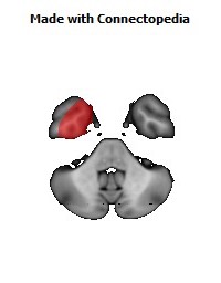


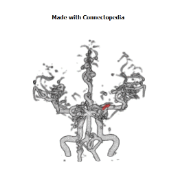

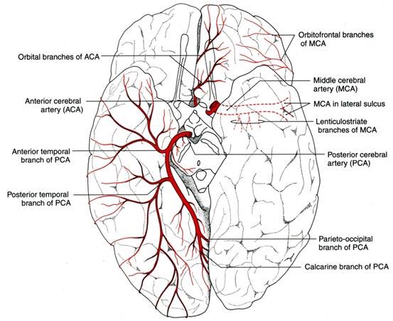
The posterior cerebral artery (PCA) is one of a pair of blood vessels that supply oxygenated blood to the posterior aspect of the brain (occipital lobe) in human anatomy. It arises near the intersection of the posterior communicating artery and the basilar artery and connects with the ipsilateral middle cerebral artery (MCA) and internal carotid artery via the posterior communicating artery (PCommA).
Origin
The development of the PCA in fetal brain comes relatively late and arises from the fusion of several embryonic vessels near the caudal end of the PCommA supplying the mesencephalon and diencephalon of the fetus. The PCA begins as such, as a continuation of the PCommA in the fetus with only 10–30% of fetuses having a prominent basilar origin.
The fetal carotid origin of the PCA usually regresses as the vertebral and basilar arteries develop with the PCommA reducing is size. In most adults, the PCA sources from the anterior portion of the basilar artery. Only about 19% of adults retain PCommA dominance of the PCA with 72% having dominant basilar origin, and the rest being equal prominence or exclusive sources for suppyling the PCA.
Structure
The branches of the posterior cerebral artery are divided into two sets, ganglionic and cortical:
Central branches
Also known as the perforating branches:
- Thalamoperforating and thalamogeniculate or postero-medial ganglionic branches: a group of small arteries which arise at the commencement of the posterior cerebral artery: these, with similar branches from the posterior communicating, pierce the posterior perforated substance, and supply the medial surfaces of the thalami and the walls of the third ventricle.
- Peduncular perforating or postero-lateral ganglionic branches: small arteries which arise from the posterior cerebral artery after it has turned around the cerebral peduncle; they supply a considerable portion of the thalamus.
Choroidal branches
See also: Anterior choroidal artery
- Medial posterior choroidal branches: run forward beneath the splenium of the corpus callosum, and supply the tela chorioidea of the third ventricle and the choroid plexus.
- Lateral posterior choroidal branches: small branches to the cerebral peduncle, fornix, thalamus, and the caudate nucleus.
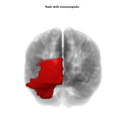
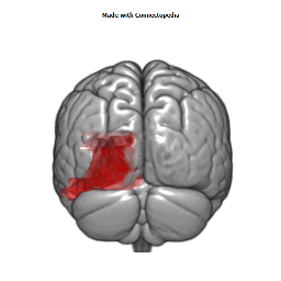
Cortical branches
The cortical branches are:
- Anterior temporal, distributed to the uncus and the anterior part of the fusiform gyrus
- Posterior temporal, to the fusiform and the inferior temporal gyri
- Lateral occipital, which branches into the anterior, middle and posterior inferior temporal arteries
- Medial occipital, which branches into the:
- Calcarine, to the cuneus and gyrus lingualis and the back part of the convex surface of the occipital lobe
- Parieto-occipital, to the cuneus and the precuneus
- Splenial, or the posterior pericallosal branch, sometimes anastamoses with the anterior cerebral artery (ACA), and may not be present if the ACA wraps around the corpus callosum
Clinical relevance
Stroke
- Contralateral loss of pain and temperature sensations.
- Visual field defects (contralateral hemianopia with macular sparing).
- Prosopagnosia with bilateral obstruction of the lingual and fusiform gyri.
- Superior Alternating Syndrome (Weber's syndrome)
- Contralateral deficits of facial nerve (only lower face, upper face receives bilateral input), vagus nerve and hypoglossal nerve
- Ipsilateral deficit of oculomotor nerve
- Horner's Syndrome
Signs and symptoms:
Structures involved
Peripheral territory (Cortical branches)
- Homonymous hemianopia (often upper quadrantic): Calcarine cortex or optic radiation nearby.
- Bilateral homonymous hemianopia, cortical blindness, awareness or denial of blindness; tactile naming, achromatopia (color blindness), failure to see to-and-fro movements, inability to perceive objects not centrally located, apraxia of ocular movements, inability to count or enumerate objects, tendency to run into things that the patient sees and tries to avoid: Bilateral occipital lobe with possibly the parietal lobe involved.
- Verbal dyslexia without agraphia, color anomia: Dominant calcarine lesion and posterior part of corpus callosum.
- Memory defect: Hippocampal lesion bilaterally or on the dominant side only.
- Topographic disorientation and prosopagnosia: Usually with lesions of nondominant, calcarine, and lingual gyrus.
- Simultanagnosia, hemivisual neglect: Dominant visual cortex, contralateral hemisphere.
- Unformed visual hallucinations, peduncular hallucinosis, metamorphopsia, teleopsia, illusory visual spread, palinopsia, distortion of outlines, central photophobia: Calcarine cortex.
- Complex hallucinations: Usually nondominant hemisphere.
Central territory (Ganglionic branches)
- Thalamic syndrome: sensory loss (all modalities), spontaneous pain and dysesthesias, choreoathetosis, intention tremor, spasms of hand, mild hemiparesis, contralateral hemianaethesia: Posteroventral nucleus of thalamus; involvement of the adjacent subthalamus body or its afferent tracts.
- Thalamoperforate syndrome: crossed cerebellar ataxia with ipsilateral third nerve palsy (Claude's syndrome): Dentatothalamic tract and issuing third nerve.
- Weber's syndrome: third nerve palsy and contralateral hemiplegia: Third nerve and cerebral peduncle.
- Contralateral hemiplegia: Cerebral peduncle.
- Paralysis or paresis of vertical eye movement, skew deviation, sluggish pupillary responses to light, slight miosis and ptosis (retraction nystagmus and "tucking" of the eyelids may be associated): Supranuclear fibers to third nerve, interstitial nucleus of Cajal, nucleus of Darkschewitsch, and posterior commissure.
- Contralateral rhythmic, ataxic action tremor; rhythmic postural or "holding" tremor (rubral tremor): Dentatothalamic tract.