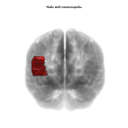


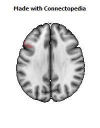
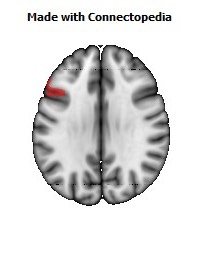
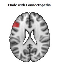
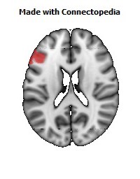
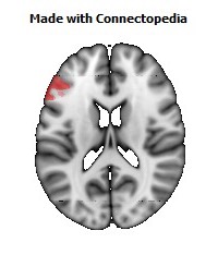
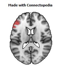
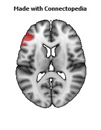
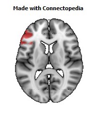
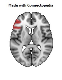
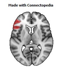
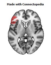
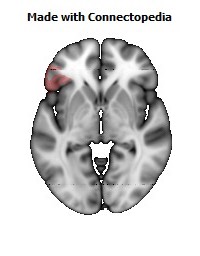


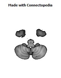


The inferior frontal gyrus is a gyrus of the frontal lobe (the yellow area of the human brain image to the right). It is labelled gyrus frontalis inferior, its Latin name. In the yellow area, its superior border is the inferior frontal sulcus (which divides it from the gyrus frontalis medius in the yellow area), its inferior border the lateral fissure (which divides it from the gyrus temporalis superior in the green area), and its posterior border is the inferior precentral sulcus (in the yellow area). Above it is the middle frontal gyrus (the gyrus frontalis medius), behind it the precentral gyrus (the gyrus praecentralis), both gyri in the yellow area of the image.
The inferior frontal gyrus, like the middle frontal gyrus and the superior frontal gyrus, is more of a region than a true gyrus.
Structure
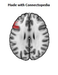
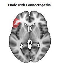
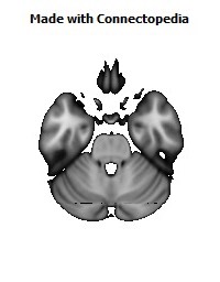
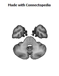
The inferior frontal gyrus can be subdivided into the following macroanatomical structures, shown in yellow in the top image, just below the label gyrus frontalis inferior:
• Pars opercularis (cortex posterior to the ascending ramus of the lateral fissure)
• Pars triangularis (cortex between the ascending ramus and the horizontal ramus of the lateral fissure)
• Pars orbitalis (cortex inferior and anterior to the horizontal ramus of the lateral fissure)
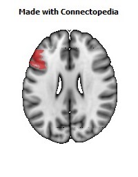
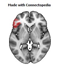
The inferior frontal gyrus includes the following cytoarchitectonic areas:
• Brodmann area 44
• Brodmann area 45
• Brodmann area 47
• cytoarchitectonic areas of the deep frontal operculum
The cytoarchitectonic areas very roughly correspond to the following macroanatomic structures: Brodmann area 44 to Pars opercularis, Brodmann area 45 to Pars triangularis, and Brodmann area 47 to Pars orbitalis. Brodmann area 44 corresponds to Broca's area (sometimes Broca's area is taken to encompass Brodmann's areas 44 and 45) — for the dominant hemisphere of the brain.
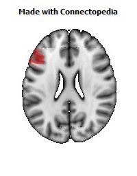
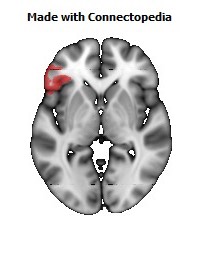
Function
The right inferior frontal gyrus (IFG) has been typically implicated in go/no go tasks. In such a task, the participant has to inhibit a prepotent response (for instance stop pressing a button when a red signal appears). It seems that the same area is also implicated in risk aversion: a study found that higher risk aversion correlated with higher activity at IFG. This might be explained as an inhibition signal to accept a risky option. Disruption of activity of this area with Transcranial Magnetic Stimulation or Direct Current Stimulation (tDCS) indeed leads to change in risk attitudes, as behaviorally demonstrated by choices over risky outcomes. The left IFG is extremely important for language production and verb comprehension (left side corresponds to the 'dominant side', for right-handed persons). Commonly known as "Broca's Area", persons with damage in this region often have non-fluent aphasia. With non-fluent aphasia, speech is notably difficult to produce but the speech that is produced is high in content compared to Wernicke's aphasiacs (persons with damage to the posterior language regions) whose aphasia is considered "fluent" yet rambling.