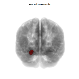

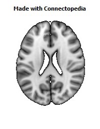
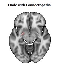
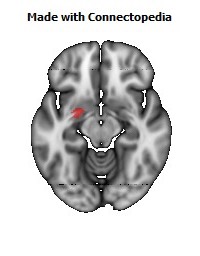
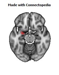
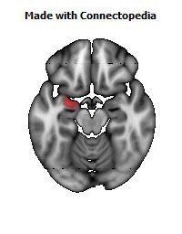
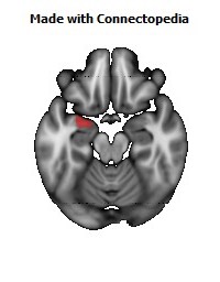
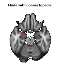
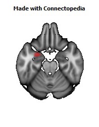
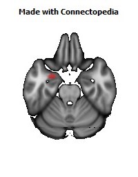
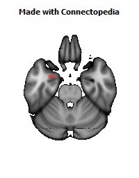
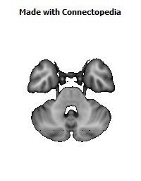
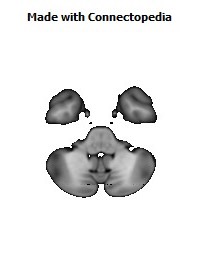


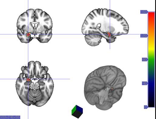
The amygdalae (singular: amygdala; /əˈmɪɡdələ/; also corpus amygdaloideum; Latin, from Greek ἀμυγδαλή, amygdalē, 'almond', 'tonsil', listed in the Gray's Anatomy textbook as the nucleus amygdalæ, are almond-shaped groups of nuclei located deep and medially within the temporal lobes of the brain in complex vertebrates, including humans. Shown in research to perform a primary role in the processing of memory, decision-making, and emotional reactions, the amygdalae are considered part of the limbic system.
Structure
The regions described as amygdala nuclei encompass several structures with distinct connectional and functional characteristics in humans and other animals. Among these nuclei are the basolateral complex, the cortical nucleus, the medial nucleus, the central nucleus, and the intercalated cell clusters (ITCs). The basolateral complex can be further subdivided into the lateral, the basal, and the accessory basal nuclei.
Anatomically, the amygdala and more particularly, its central and medial nuclei, have sometimes been classified as a part of the basal ganglia.
Hemispheric specializations
There are functional differences between the right and left amygdala. In one study, electrical stimulations of the right amygdala induced negative emotions, especially fear and sadness. In contrast, stimulation of the left amygdala was able to induce either pleasant (happiness) or unpleasant (fear, anxiety, sadness) emotions. Other evidence suggests that the left amygdala plays a role in the brain's reward system.
Sex differences
The amygdala is one of the most well understood brain regions with regard to differences between the sexes. Larger male than female amygdalae have been demonstrated in children ages 7–11, in adult humans, and in adult rats.
In addition to size, other differences between men and women exist with regards to the amygdala. Subjects' amygdala activation was observed when watching a horror film. The results of the study showed a different lateralization of the amygdala in men and women. Enhanced memory for the film was related to enhanced activity of the left, but not the right, amygdala in women, whereas it was related to enhanced activity of the right, but not the left, amygdala in men. One study found evidence that on average, women tend to retain stronger memories for emotional events than men.
The right amygdala is also linked with taking action as well as being linked to negative emotions, which may help explain why males tend to respond to emotionally stressful stimuli physically. The left amygdala allows for the recall of details, but it also results in more thought rather than action in response to emotionally stressful stimuli, which may explain the lack of physical response in women.
Function
Connections
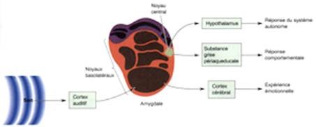
The amygdala sends projections to the hypothalamus, the dorsomedial thalamus, the thalamic reticular nucleus, the nuclei of the trigeminal nerve and the facial nerve, the ventral tegmental area, the locus coeruleus, and the laterodorsal tegmental nucleus.
The medial nucleus is involved in the sense of smell and pheromone-processing. It receives input from the olfactory bulb and olfactory cortex. The lateral amygdalae, which send impulses to the rest of the basolateral complexes and to the centromedial nuclei, receive input from the sensory systems. The centromedial nuclei are the main outputs for the basolateral complexes, and are involved in emotional arousal in rats and cats.
Emotional learning
In complex vertebrates, including humans, the amygdalae perform primary roles in the formation and storage of memories associated with emotional events. Research indicates that, during fear conditioning, sensory stimuli reach the basolateral complexes of the amygdalae, particularly the lateral nuclei, where they form associations with memories of the stimuli. The association between stimuli and the aversive events they predict may be mediated by long-term potentiation, a sustained enhancement of signaling between affected neurons. There have been studies that show that damage to the amygdala can interfere with memory that is strengthened by emotion. One study examined a patient with bilateral degeneration of the amygdala. He was told a violent story accompanied by matching pictures and was observed based on how much he could recall from the story. The patient had less recollection of the story than patients without amgydala damage, showing that the amgydala has a strong connection with emotional learning.
Memories of emotional experiences imprinted in reactions of synapses in the lateral nuclei elicit fear behavior through neuronal connections with the central nucleus of the amygdalae and the bed nuclei of the stria terminalis (BNST). The axon terminals from sensory neurons form synapses with dendritic spines on neurons from the central nucleus. The central nuclei are involved in the genesis of many fear responses such as defensive behavior (freezing or escape responses), autonomic nervous system responses (changes in blood pressure and heart rate/tachycardia), neuroendocrine responses (stress-hormone release), etc. Damage to the amygdalae impairs both the acquisition and expression of Pavlovian fear conditioning, a form of classical conditioning of emotional responses.
The amygdalae are also involved in appetitive (positive) conditioning. It seems that distinct neurons respond to positive and negative stimuli, but there is no clustering of these distinct neurons into clear anatomical nuclei. However, lesions of the central nucleus in the amygdala have been shown to reduce appetitive learning in rats. Lesions of the basolateral regions do not exhibit the same effect. Research like this indicates that different nuclei within the amygdala have different functions in appetitive conditioning.
Memory modulation
The amygdala is also involved in the modulation of memory consolidation. Following any learning event, the long-term memory for the event is not formed instantaneously. Rather, information regarding the event is slowly assimilated into long-term (potentially lifelong) storage over time, possibly via long-term potentiation. Recent studies suggest that, while the amygdala is not itself a long-term memory storage site, and learning can occur without it, one of its roles is to regulate memory consolidation in other brain regions. Also, fear conditioning, a type of memory that is impaired following amygdala damage, is mediated in part by long-term potentiation.
During the consolidation period, the memory can be modulated. In particular, it appears that emotional arousal following the learning event influences the strength of the subsequent memory for that event. Greater emotional arousal following a learning event enhances a person's retention of that event. Experiments have shown that administration of stress hormones to mice immediately after they learn something enhances their retention when they are tested two days later.
The amygdala, especially the basolateral nuclei, are involved in mediating the effects of emotional arousal on the strength of the memory for the event, as shown by many laboratories including that of James McGaugh. These laboratories have trained animals on a variety of learning tasks and found that drugs injected into the amygdala after training affect the animals' subsequent retention of the task. These tasks include basic classical conditioning tasks such as inhibitory avoidance, where a rat learns to associate a mild footshock with a particular compartment of an apparatus, and more complex tasks such as spatial or cued water maze, where a rat learns to swim to a platform to escape the water. If a drug that activates the amygdalae is injected into the amygdalae, the animals had better memory for the training in the task. If a drug that inactivates the amygdalae is injected, the animals had impaired memory for the task.
Buddhist monks who do compassion meditation have been shown to modulate their amygdala, along with their temporoparietal junction and insula, during their practice. In an fMRI study, more intensive insula activity was found in expert meditators than in novices. Increased activity in the amygdala following compassion-oriented meditation may contribute to social connectedness.
Amygdala activity at the time of encoding information correlates with retention for that information. However, this correlation depends on the relative "emotionalness" of the information. More emotionally-arousing information increases amygdalar activity, and that activity correlates with retention. Amygdala neurons show various types of oscillation during emotional arousal, such as theta activity. These synchronized neuronal events could promote synaptic plasticity (which is involved in memory retention) by increasing interactions between neocortical storage sites and temporal lobe structures involved in declarative memory.
Research using Rorschach test blot 03 finds that the number of unique responses to this random figure links to larger sized amygdalae. The researchers note, "Since previous reports have indicated that unique responses were observed at higher frequency in the artistic population than in the nonartistic normal population, this positive correlation suggests that amygdalar enlargement in the normal population might be related to creative mental activity."
Neuropsychological correlates of amygdala activity
Early research on primates provided explanations as to the functions of the amygdala, as well as a basis for further research. As early as 1888, rhesus monkeys with a lesioned temporal cortex (including the amygdala) were observed to have significant social and emotional deficits. Heinrich Klüver and Paul Bucy later expanded upon this same observation by showing that large lesions to the anterior temporal lobe produced noticeable changes, including overreaction to all objects, hypoemotionality, loss of fear, hypersexuality, and hyperorality, a condition in which inappropriate objects are placed in the mouth. Some monkeys also displayed an inability to recognize familiar objects and would approach animate and inanimate objects indiscriminately, exhibiting a loss of fear towards the experimenters. This behavioral disorder was later named Klüver-Bucy syndrome accordingly, and later research proved it was specifically due to amygdala lesions. Monkey mothers who had amygdala damage showed a reduction in maternal behaviors towards their infants, often physically abusing or neglecting them. In 1981, researchers found that selective radio frequency lesions of the whole amygdala caused Klüver-Bucy Syndrome.
With advances in neuroimaging technology such as MRI, neuroscientists have made significant findings concerning the amygdala in the human brain. A variety of data shows the amygdala has a substantial role in mental states, and is related to many psychological disorders. Some studies have shown children with anxiety disorders tend to have a smaller left amygdala. In the majority of the cases, there was an association between an increase in the size of the left amygdala with the use of SSRI's (antidepressant medication) or psychotherapy. The left amygdala has been linked to social anxiety, obsessive and compulsive disorders, and post traumatic stress, as well as more broadly to separation and general anxiety. In a 2003 study, subjects with borderline personality disorder showed significantly greater left amygdala activity than normal control subjects. Some borderline patients even had difficulties classifying neutral faces or saw them as threatening. Individuals with psychopathy show reduced autonomic responses, relative to comparison individuals, to instructed fear cues. In 2006, researchers observed hyperactivity in the amygdala when patients were shown threatening faces or confronted with frightening situations. Patients with more severe social phobia showed a correlation with increased response in the amygdala. Similarly, depressed patients showed exaggerated left amygdala activity when interpreting emotions for all faces, and especially for fearful faces. Interestingly, this hyperactivity was normalized when patients were administered antidepressant medication. By contrast, the amygdala has been observed to respond differently in people with bipolar disorder. A 2003 study found that adult and adolescent bipolar patients tended to have considerably smaller amygdala volumes and somewhat smaller hippocampal volumes. Many studies have focused on the connections between the amygdala and autism.
Studies in 2004 and 2006 showed that normal subjects exposed to images of frightened faces or faces of people from another race will show increased activity of the amygdala, even if that exposure is subliminal. However, the amygdala is not necessary for the processing of fear-related stimuli, since persons in whom it is bilaterally damaged show rapid reactions to fearful faces, even in the absence of a functional amygdala.
Recent research suggests that parasites, in particular toxoplasma, form cysts in the brain of rats, often taking up residence in the amygdala. This may provide clues as to how specific parasites may contribute to the development of disorders, including paranoia.
Future studies have been proposed to address the role of the amygdala in positive emotions, and the ways in which the amygdala networks with other brain regions.
Sexual orientation
Recent studies have suggested possible correlations between brain structure, including differences in hemispheric ratios and connection patterns in the amygdala, and sexual orientation. Homosexual men tend to exhibit more female-like patterns in the amygdala than heterosexual males do, just as homosexual females tend to show more male-like patterns in the amygdala than heterosexual women do. It was observed that amygdala connections were more widespread from the left amygdala in homosexual males, as is also found in heterosexual females. Amygdala connections were more widespread from the right amygdala in homosexual females, as in heterosexual males. Current evidence suggests that formation of an individual's sexual orientation occurs during fetal and neonatal development.
Social interaction
Amygdala volume correlates positively with both the size (the number of contacts a person has) and the complexity (the number of different groups to which a person belongs) of social networks. Individuals with larger amygdalae had larger and more complex social networks. They were also better able to make accurate social judgments about other persons' faces. It is hypothesized that larger amygdalae allow for greater emotional intelligence, enabling greater societal integration and cooperation with others.
The amygdala processes reactions to violations concerning personal space. These reactions are absent in persons in whom the amygdala is damaged bilaterally. Furthermore, the amygdala is found to be activated in fMRI when people observe that others are physically close to them, such as when a person being scanned knows that an experimenter is standing immediately next to the scanner, versus standing at a distance.
Aggression
Animal studies have shown that stimulating the amygdala appears to increase both sexual and aggressive behavior. Likewise, studies using brain lesions have shown that harm to the amygdala may produce the opposite effect. Thus, it appears that this part of the brain may play a role in the display and modulation of aggression.
Fear
There are cases of human patients with focal bilateral amygdala lesions, due to the rare genetic condition Urbach-Wiethe disease. Such patients fail to exhibit fear-related behaviors, leading one to be dubbed the "woman with no fear". This finding reinforces the conclusion that the amygdala "plays a pivotal role in triggering a state of fear".
Alcoholism and binge drinking
The amygdala appears to play a role in binge drinking, being damaged by repeated episodes of intoxication and withdrawal. Alcoholism is associated with dampened activation in brain networks responsible for emotional processing, including the amygdala. Protein kinase C-epsilon in the amygdala is important for regulating behavioral responses to morphine, ethanol, and controlling anxiety-like behavior. The protein is involved in controlling the function of other proteins and plays a role in development of the ability to consume a large amount of ethanol.
Anxiety
There may also be a link between the amygdala and anxiety. In particular, there is a higher prevalence of females that are affected by anxiety disorders. In an experiment, degu pups were removed from their mother but allowed to hear her call. In response, the males produced increased serotonin receptors in the amygdala but females lost them. This led to the males being less affected by the stressful situation.
Posttraumatic Stress Disorder
There seems to be a connection with the amygdala and how the brain processes posttraumatic stress disorder. Multiple studies have found that the amgydala may be responsible for the emotional reactions of PTSD patients. One study in specific found that when PTSD patients are shown pictures of faces with fearful expressions, their amygdalas tended to have a higher activation than someone without PTSD.