
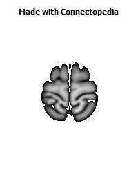
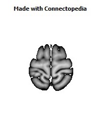
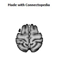
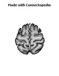
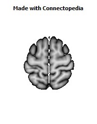
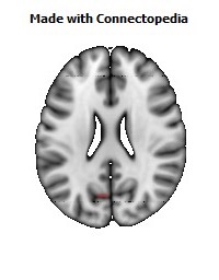
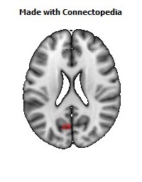
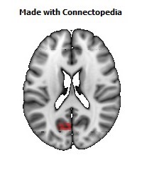
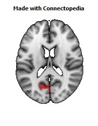
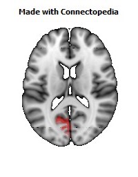
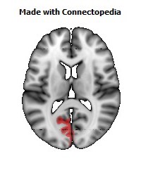
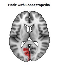
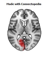
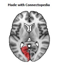
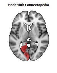
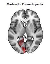
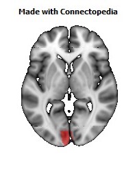
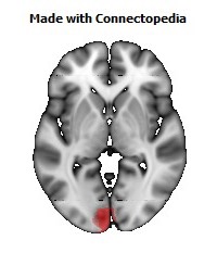
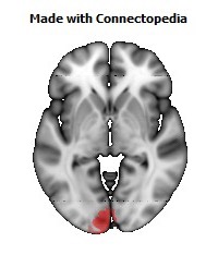
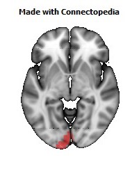
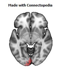
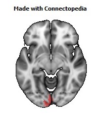
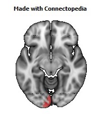
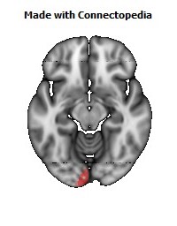
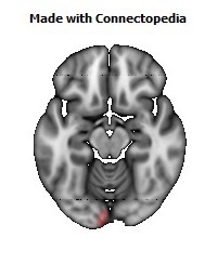
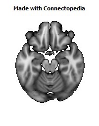
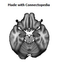

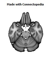
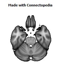
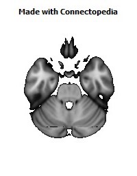
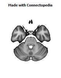
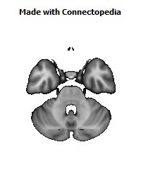
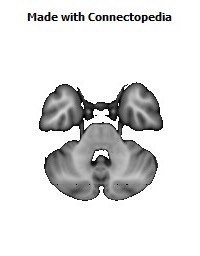
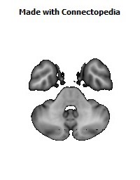
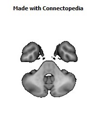
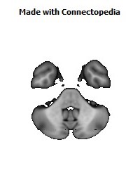
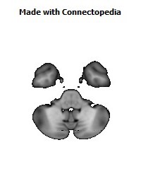


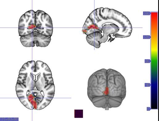
The calcarine fissure (or calcarine sulcus) is an anatomical landmark located at the caudal end of the medial surface of the brain. Its name comes from the Latin "calcar" meaning "spur." It is a complete sulcus.
Anatomy
The calcarine sulcus begins near the occipital pole in two converging rami and runs forward to a point a little below the splenium of the corpus callosum, where it is joined at an acute angle by the medial part of the parieto-occipital sulcus. The anterior part of this fissure gives rise to the prominence of the calcar avis in the posterior cornu of the lateral ventricle.
Function
The calcarine sulcus is where the primary visual cortex (V1) is concentrated. The central visual field is located in the posterior portion of the calcarine sulcus and the peripheral visual field in the anterior portion.
Visual cortex
The visual cortex of the brain is the part of the cerebral cortex responsible for processing visual information. It is located in the occipital lobe, at the back of the brain. The visual cortex is made up of Brodmann area 17, (the primary visual cortex), Brodmann area 18 and Brodmann area 19, the extrastriate cortical areas.
The primary visual cortex is also known as V1, visual area one, and also as the striate cortex. The extrastriate areas are referred to as V2,V3, V4, and V5, visual areas two, three, four and five. There is a visual cortex in each hemisphere of the brain. The left hemisphere visual cortex receives signals from the right visual field and the right visual cortex from the left visual field.
The body of this article describes the primate (especially, human) visual cortex.
Introduction
The primary visual cortex, V1, is the koniocortex (sensory type) located in and around the calcarine fissure in the occipital lobe. Each hemisphere's V1 receives information directly from its ipsilateral lateral geniculate nucleus.
Each V1 transmits information to two primary pathways, called the dorsal stream and the ventral stream:
• The dorsal stream begins with V1, goes through Visual area V2, then to the dorsomedial area and Visual area MT (also known as V5) and to the posterior parietal cortex. The dorsal stream, sometimes called the "Where Pathway" or "How Pathway", is associated with motion, representation of object locations, and control of the eyes and arms, especially when visual information is used to guide saccades or reaching.
• The ventral stream begins with V1, goes through visual area V2, then through visual area V4, and to the inferior temporal cortex. The ventral stream, sometimes called the "What Pathway", is associated with form recognition and object representation. It is also associated with storage of long-term memory.
The what vs. where account of the ventral/dorsal pathways was first described by Ungerleider and Mishkin. More recently, Goodale and Milner extended these ideas and suggested that the ventral stream is critical for visual perception whereas the dorsal stream mediates the visual control of skilled actions. Although these ideas are contentious among some vision scientists and psychologists, the proposed division of labor between vision-for-perception and vision-for-action is supported by a broad range of findings from neuropsychology, neurophysiology, and neuroimaging. Some of the most controversial evidence comes from studies of visuomotor behaviour in normal observers. For example, it has been shown that visual illusions such as the Ebbinghaus illusion distort judgements of a perceptual nature, but when the subject responds with an action, such as grasping, no distortion occurs. Other work suggests that both the action and perception systems are equally fooled by such illusions. More recent studies, however, provide strong support for the idea that skilled actions such as grasping are not affected by pictorial illusions. and suggest that the action/perception dissociation is a useful way to characterize the functional division of labor between the dorsal and ventral visual pathways in the cerebral cortex.
Neurons in the visual cortex fire action potentials when visual stimuli appear within their receptive field. By definition, the receptive field is the region within the entire visual field that elicits an action potential. But, for any given neuron, it may respond best to a subset of stimuli within its receptive field. This property is called neuronal tuning. In the earlier visual areas, neurons have simpler tuning. For example, a neuron in V1 may fire to any vertical stimulus in its receptive field. In the higher visual areas, neurons have complex tuning. For example, in the inferior temporal cortex (IT), a neuron may fire only when a certain face appears in its receptive field.
The visual cortex receives its blood supply primarily from the calcarine branch of the posterior cerebral artery.
Current research
Research on the primary visual cortex can involve recording action potentials from electrodes within the brain of cats, ferrets, rats, mice, or monkeys, or through recording intrinsic optical signals from animals or EEG, MEG, or fMRI signals from human and monkey V1.
One recent discovery concerning the human V1 is that signals measured by fMRI show very large attentional modulation. This result is consistent with another recent electrophysiology study. The study found that although V1 processes visual information before V2 and V4, the attentional modulation occurs in V4 followed by V2 and then finally in V1 suggesting attentional modulation occurs through feedback from higher-level visual areas to lower-level visual areas.
Other current work on V1 seeks to fully characterize its tuning properties, and to use it as a model area for the canonical cortical circuit.
Lesions to primary visual cortex usually lead to a scotoma, or hole in the visual field. Note that patients with scotomas are often able to make use of visual information presented to their scotomas, despite being unable to consciously perceive it. This phenomenon, called blindsight, is widely studied by scientists interested in the neural correlates of consciousness.
Concurrent research work in the domain of computer vision, addressing automatic object recognition by computer algorithm, has looked to draw inspiration from current understanding in this biological domain.
Primary visual cortex (V1)
The primary visual cortex is the best-studied visual area in the brain. In all mammals studied, it is located in the posterior pole of the occipital cortex (the occipital cortex is responsible for processing visual stimuli). It is the simplest, earliest cortical visual area. It is highly specialized for processing information about static and moving objects and is excellent in pattern recognition.
The functionally defined primary visual cortex is approximately equivalent to the anatomically defined striate cortex. The name "striate cortex" is derived from the line of Gennari, a distinctive stripe visible to the naked eye that represents myelinated axons from the lateral geniculate body terminating in layer 4 of the gray matter.
The primary visual cortex is divided into six functionally distinct layers, labeled 1 through 6. Layer 4, which receives most visual input from the lateral geniculate nucleus (LGN), is further divided into 4 layers, labelled 4A, 4B, 4Cα, and 4Cβ. Sublamina 4Cα receives most magnocellular input from the LGN, while layer 4Cβ receives input from parvocellular pathways.
The average number of neurons in the adult human primary visual cortex, in each hemisphere, has been estimated at around 140 million.
Function
V1 has a very well-defined map of the spatial information in vision. For example, in humans, the upper bank of the calcarine sulcus responds strongly to the lower half of visual field (below the center), and the lower bank of the calcarine to the upper half of visual field. In concept, this retinotopic mapping is a transformation of the visual image from retina to V1. The correspondence between a given location in V1 and in the subjective visual field is very precise: even the blind spots are mapped into V1. In terms of evolution, this correspondence is very basic and found in most animals that possess a V1. In human and animals with a fovea in the retina, a large portion of V1 is mapped to the small, central portion of visual field, a phenomenon known as cortical magnification. Perhaps for the purpose of accurate spatial encoding, neurons in V1 have the smallest receptive field size of any visual cortex microscopic regions.
The tuning properties of V1 neurons (what the neurons respond to) differ greatly over time. Early in time (40 ms and further) individual V1 neurons have strong tuning to a small set of stimuli. That is, the neuronal responses can discriminate small changes in visual orientations, spatial frequencies and colors. Furthermore, individual V1 neurons in human and animals with binocular vision have ocular dominance, namely tuning to one of the two eyes. In V1, and primary sensory cortex in general, neurons with similar tuning properties tend to cluster together as cortical columns. David Hubel and Torsten Wiesel proposed the classic ice-cube organization model of cortical columns for two tuning properties: ocular dominance and orientation. However, this model cannot accommodate the color, spatial frequency and many other features to which neurons are tuned. The exact organization of all these cortical columns within V1 remains a hot topic of current research.
Current consensus seems to be that early responses of V1 neurons consists of tiled sets of selective spatiotemporal filters. In the spatial domain, the functioning of V1 can be thought of as similar to many spatially local, complex Fourier transforms, or more accurately, Gabor transforms. In theory, these filters together can carry out neuronal processing of spatial frequency, orientation, motion, direction, speed (thus temporal frequency), and many other spatiotemporal features. Experiments of neurons substantiate these theories, but also raise new questions.
Later in time (after 100 ms), neurons in V1 are also sensitive to the more global organisation of the scene (Lamme & Roelfsema, 2000). These response properties probably stem from recurrent processing (the influence of higher-tier cortical areas on lower-tier cortical areas) and lateral connections from pyramidal neurons (Hupe et al. 1998). While feedforward connections are mainly driving, feedback connections are mostly modulatory in their effects (Angelucci et al., 2003; Hupe et al., 2001). Evidence shows that feedback originating in higher-level areas such as V4, IT, or MT, with bigger and more complex receptive fields, can modify and shape V1 responses, accounting for contextual or extra-classical receptive field effects (Guo et al., 2007; Huang et al., 2007; Sillito et al., 2006).
The visual information relayed to V1 is not coded in terms of spatial (or optical) imagery but rather as the local contrast. As an example, for an image comprising half side black and half side white, the divide line between black and white has strongest local contrast and is encoded, while few neurons code the brightness information (black or white per se). As information is further relayed to subsequent visual areas, it is coded as increasingly non-local frequency/phase signals. Note that, at these early stages of cortical visual processing, spatial location of visual information is well preserved amid the local contrast encoding.
V2
Visual area V2, also called prestriate cortex, is the second major area in the visual cortex, and the first region within the visual association area. It receives strong feedforward connections from V1 (direct and via the pulvinar) and sends strong connections to V3, V4, and V5. It also sends strong feedback connections to V1.
In terms of anatomy, V2 is split into four quadrants, a dorsal and ventral representation in the left and the right hemispheres. Together, these four regions provide a complete map of the visual world. V2 has many properties in common with V1: Cells are tuned to simple properties such as orientation, spatial frequency, and color. The responses of many V2 neurons are also modulated by more complex properties, such as the orientation of illusory contours, binocular disparity, and whether the stimulus is part of the figure or the ground (Qiu and von der Heydt, 2005). Recent research has shown that V2 cells show a small amount of attentional modulation (more than V1, less than V4), are tuned for moderately complex patterns, and may be driven by multiple orientations at different subregions within a single receptive field.
It is argued that the entire ventral visual-to-hippocampal stream is important for visual memory. This theory, unlike the dominant one, predicts that object-recognition memory (ORM) alterations could result from the manipulation in V2, an area that is highly interconnected within the ventral stream of visual cortices. In the monkey brain, this area receives strong feedforward connections from the primary visual cortex (V1) and sends strong projections to other secondary visual cortices (V3, V4, and V5). Most of the neurons of this area are tuned to simple visual characteristics such as orientation, spatial frequency, size, color, and shape. V2 cells also respond to various complex shape characteristics, such as the orientation of illusory contours and whether the stimulus is part of the figure or the ground. Anatomical studies implicate layer 3 of area V2 in visual-information processing. In contrast to layer 3, layer 6 of the visual cortex is composed of many types of neurons, and their response to visual stimuli is more complex.
In a recent study, the Layer 6 cells of the V2 cortex were found to play a very important role in the storage of Object Recognition Memory as well as the conversion of short-term object memories into long-term memories.
Third visual complex, including area V3
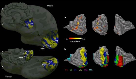
The term third visual complex refers to the region of cortex located immediately in front of V2, which includes the region named visual area V3 in humans. The "complex" nomenclature is justified by the fact that some controversy still exists regarding the exact extent of area V3, with some researchers proposing that the cortex located in front of V2 may include two or three functional subdivisions. For example, David Van Essen and others (1986) have proposed the existence of a "dorsal V3" in the upper part of the cerebral hemisphere, which is distinct from the "ventral V3" (or ventral posterior area, VP) located in the lower part of the brain. Dorsal and ventral V3 have distinct connections with other parts of the brain, appear different in sections stained with a variety of methods, and contain neurons that respond to different combinations of visual stimulus (for example, colour-selective neurons are more common in the ventral V3). Additional subdivisions, including V3A and V3B have also been reported in humans. These subdivisions are located near dorsal V3, but do not adjoin V2.
Dorsal V3 is normally considered to be part of the dorsal stream, receiving inputs from V2 and from the primary visual area and projecting to the posterior parietal cortex. It may be anatomically located in Brodmann area 19. Braddick using fMRI has suggested that area V3/V3A may play a role in the processing of global motion. Other studies prefer to consider dorsal V3 as part of a larger area, named the dorsomedial area (DM), which contains a representation of the entire visual field. Neurons in area DM respond to coherent motion of large patterns covering extensive portions of the visual field (Lui and collaborators, 2006).
Ventral V3 (VP), has much weaker connections from the primary visual area, and stronger connections with the inferior temporal cortex. While earlier studies proposed that VP contained a representation of only the upper part of the visual field (above the point of fixation), more recent work indicates that this area is more extensive than previously appreciated, and like other visual areas it may contain a complete visual representation. The revised, more extensive VP is referred to as the ventrolateral posterior area (VLP) by Rosa and Tweedale.
V4
Visual area V4 is one of the visual areas in the extrastriate visual cortex. In macaques, it is located anterior to V2 and posterior to posterior inferotemporal area (PIT). It comprises at least four regions (left and right V4d, left and right V4v), and some groups report that it contains rostral and caudal subdivisions as well. It is unknown what the human homologue of V4 is, and this issue is currently the subject of much scrutiny.
V4 is the third cortical area in the ventral stream, receiving strong feedforward input from V2 and sending strong connections to the PIT. It also receives direct inputs from V1, especially for central space. In addition, it has weaker connections to V5 and dorsal prelunate gyrus (DP).
V4 is the first area in the ventral stream to show strong attentional modulation. Most studies indicate that selective attention can change firing rates in V4 by about 20%. A seminal paper by Moran and Desimone characterizing these effects was the first paper to find attention effects anywhere in the visual cortex.
Like V1, V4 is tuned for orientation, spatial frequency, and color. Unlike V1, V4 is tuned for object features of intermediate complexity, like simple geometric shapes, although no one has developed a full parametric description of the tuning space for V4. Visual area V4 is not tuned for complex objects such as faces, as areas in the inferotemporal cortex are.
The firing properties of V4 were first described by Semir Zeki in the late 1970s, who also named the area. Before that, V4 was known by its anatomical description, the prelunate gyrus. Originally, Zeki argued that the purpose of V4 was to process color information. Work in the early 1980s proved that V4 was as directly involved in form recognition as earlier cortical areas. This research supported the Two Streams hypothesis, first presented by Ungerleider and Mishkin in 1982.
Recent work has shown that V4 exhibits long-term plasticity, encodes stimulus salience, is gated by signals coming from the frontal eye fields and shows changes in the spatial profile of its receptive fields with attention.
V5/MT
Visual area V5, also known as visual area MT (middle temporal), is a region of extrastriate visual cortex that is thought to play a major role in the perception of motion, the integration of local motion signals into global percepts, and the guidance of some eye movements.
Connections
MT is connected to a wide array of cortical and subcortical brain areas. Its inputs include the visual cortical areas V1, V2, and dorsal V3 (dorsomedial area), the koniocellular regions of the LGN, and the inferior pulvinar. The pattern of projections to MT changes somewhat between the representations of the foveal and peripheral visual fields, with the latter receiving inputs from areas located in the midline cortex and retrosplenial region.
A standard view is that V1 provides the "most important" input to MT. Nonetheless, several studies have demonstrated that neurons in MT are capable of responding to visual information, often in a direction-selective manner, even after V1 has been destroyed or inactivated. Moreover, research by Semir Zeki and collaborators has suggested that certain types of visual information may reach MT before it even reaches V1.
MT sends its major outputs to areas located in the cortex immediately surrounding it, including areas FST, MST, and V4t (middle temporal crescent). Other projections of MT target the eye movement-related areas of the frontal and parietal lobes (frontal eye field and lateral intraparietal area).
Function
The first studies of the electrophysiological properties of neurons in MT showed that a large portion of the cells are tuned to the speed and direction of moving visual stimuli. These results suggested that MT plays a significant role in the processing of visual motion.
Lesion studies have also supported the role of MT in motion perception and eye movements. Neuropsychological studies of a patient unable to see motion, seeing the world in a series of static "frames" instead, suggested that V5 in the primate is homologous to MT in the human.
However, since neurons in V1 are also tuned to the direction and speed of motion, these early results left open the question of precisely what MT could do that V1 could not. Much work has been carried out on this region, as it appears to integrate local visual motion signals into the global motion of complex objects. For example, lesion to the V5 leads to deficits in perceiving motion and processing of complex stimuli. It contains many neurons selective for the motion of complex visual features (line ends, corners). Microstimulation of a neuron located in the V5 affects the perception of motion. For example, if one finds a neuron with preference for upward motion, and then we use an electrode to stimulate it, the monkey becomes more likely to report 'upward' motion.
There is still much controversy over the exact form of the computations carried out in area MT and some research suggests that feature motion is in fact already available at lower levels of the visual system such as V1.
Functional organization
MT was shown to be organized in direction columns. DeAngelis argued that MT neurons were also organized based on their tuning for binocular disparity.
V6
The dorsomedial area (DM) also known as V6, appears to respond to visual stimuli associated with self-motion (Sensitivity of human visual and vestibular cortical regions to egomotion-compatible visual stimulation, Cardin and Smith, 2010) and wide-field stimulation (Wide-Field Retinotopy Defines Human Cortical Visual Area V6, Pitzalis et al. 2006). V6, is a subdivision of the visual cortex of primates first described by John Allman and Jon Kaas in 1975. V6 is located in the dorsal part of the extrastriate cortex, near the deep groove through the centre of the brain (medial longitudinal fissure), and typically also includes portions of the medial cortex, such as the parieto-occipital sulcus. DM contains a topographically organized representation of the entire field of vision. Like the middle temporal area V5, DM receives direct connections from the primary visual cortex. Also similar to V5, DM is also characterized by high myelin content, a characteristic that is usually present in brain structures involved in fast transmission of information (Rosa et al. 2005).
For many years, it was considered that DM only existed in New World monkeys. However, more recent research has suggested that DM also exists in Old World monkeys and perhaps humans. V6 is also sometimes referred to as the parietooccipital area (PO), although the correspondence is not exact (Galletti et al. 2005).
Properties
Neurons in area DM/V6 have unique response properties, including an extremely sharp selectivity for the orientation of visual contours, and preference for long, uninterrupted lines covering large parts of the visual field (Baker and collaborators, 1981; Lui and collaborators, 2006). However, in comparison with area MT, a much smaller proportion of DM cells shows selectivity for the direction of motion of visual patterns. Another notable difference is that cells in DM are attuned to low spatial frequency components of an image, and respond poorly to the motion of textured patterns such as a field of random dots. In contrast, cells in MT are often strongly responsive to such stimuli. These response properties suggest that DM and MT may work in parallel, with the former analyzing self-motion relative to the environment, and the latter analyzing the motion of individual objects relative to the background.
Recently, an area responsive to wide-angle flow fields has been identified in the human and is thought to be a homologue of macaque area V6.
Pathways
The connections and response properties of cells in DM/ V6 suggest that this area is a key node in a sub-set of the "dorsal stream", referred to by some as the "dorsomedial pathway". This pathway is likely to be important for the control of skeletomotor activity, including postural reactions and reaching movements towards objects (Galletti and collaborators, 2003). The main "feedforward" connection of DM is to the cortex immediately rostral to it, in the interface between the occipital and parietal lobes (V6A). This region has, in turn, relatively direct connections with the regions of the frontal lobe that control arm movements, including the premotor cortex.