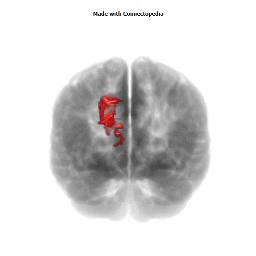

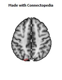
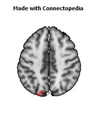
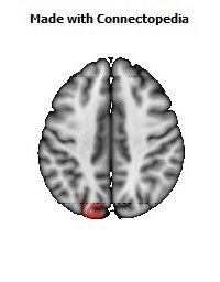
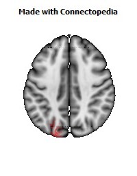
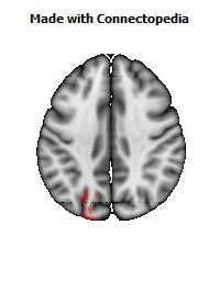
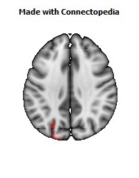
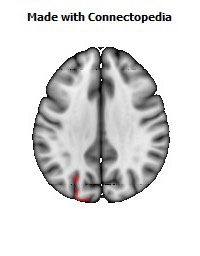
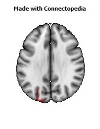
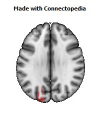
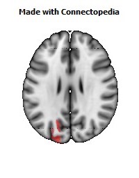
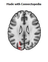
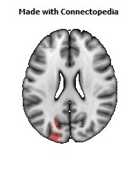
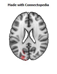
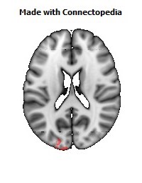
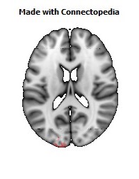
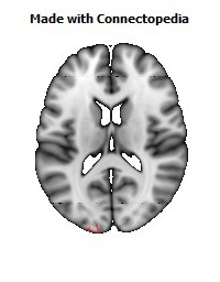
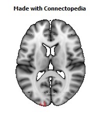
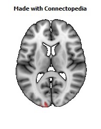
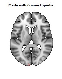
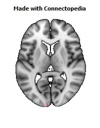
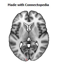
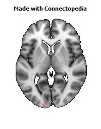
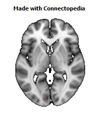
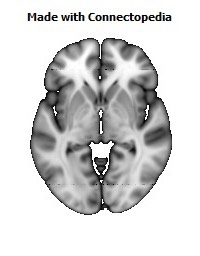
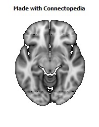
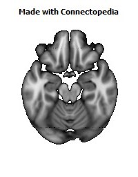
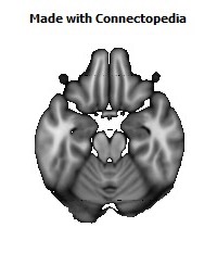

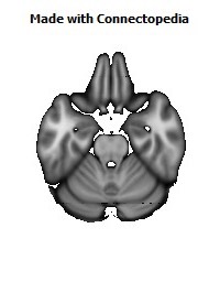
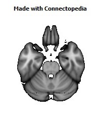
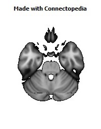
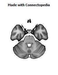
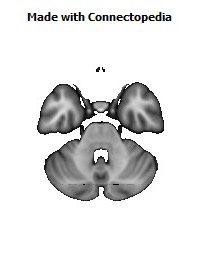
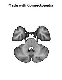
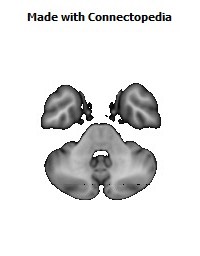
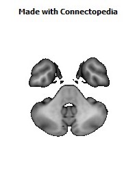
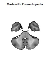
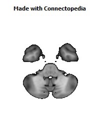
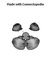
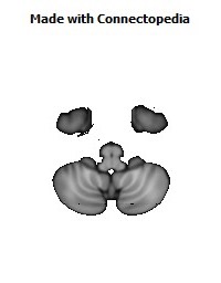


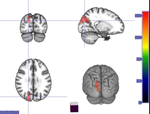
Brodmann area 19, or BA 19, is part of the occipital lobe cortex in the human brain. Along with area 18, it comprises the extrastriate (or peristriate) cortex. In humans with normal sight, extrastriate cortex is a visual association area, with feature-extracting, shape recognition, attentional, and multimodal integrating functions.
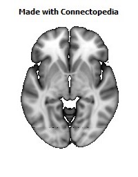
This area is also known as peristriate area 19, and it refers to a subdivision of the cytoarchitecturally defined occipital region of cerebral cortex. In the human it is located in parts of the lingual gyrus, the cuneus, the lateral occipital gyrus (H) and the superior occipital gyrus (H) of the occipital lobe where it is bounded approximately by the parieto-occipital sulcus. It is bounded on one side by the parastriate area 18, which it surrounds. It is bounded rostrally by the angular area 39 (H) and the occipitotemporal area 37 (H) (Brodmann-1909).
Function
Area 19 is a histologically delineated band anterolaterally abutting visual area 18. Single-cell electrophysiological recordings from area 19 in the cat suggest sensitivity to motion-delineated forms; recordings from primates have yielded varying results, indicating that this area may be a heterogeneous collection of visual areas, with multiple incomplete representations of the visual scene.
In humans, this band is reputed to contain regions of the visual areas designated V3, V4, V5 (also known as the middle temporal area, or MT), and V6 (also known as dorsomedial area) in the primate. Functional magnetic resonance imaging shows the existence of various retinotopic maps within area 19. In general, the diverse fields that comprise area 19 have reciprocal connections with areas 17 and 18, as well as posterior parietal and inferior temporal association areas.
Area 19 has been noted to receive inputs from the retina via the superior colliculus and pulvinar, and may contribute to the phenomenon of blindsight. In patients blind from a young age, the area has been found to be activated by somatosensory stimuli.
Because of these findings, it is thought that area 19 is the differentiation point of the two visual streams, of the 'what' and 'where' visual pathways. The dorsal region may contain motion-sensitive neurons, and ventral areas may be specialised for object recognition.
Occipital Lobe
The occipital lobe is one of the four major lobes of the cerebral cortex in the brain of mammals. The occipital lobe is the visual processing center of the mammalian brain containing most of the anatomical region of the visual cortex. The primary visual cortex is Brodmann area 17, commonly called V1 (visual one). Human V1 is located on the medial side of the occipital lobe within the calcarine sulcus; the full extent of V1 often continues onto the posterior pole of the occipital lobe. V1 is often also called striate cortex because it can be identified by a large stripe of myelin, the Stria of Gennari. Visually driven regions outside V1 are called extrastriate cortex. There are many extrastriate regions, and these are specialized for different visual tasks, such as visuospatial processing, color discrimination, and motion perception. The name derives from the overlying occipital bone, which is named from the Latin ob, behind, and caput, the head. Bilateral lesions of the occipital lobe can lead to cortical blindness.
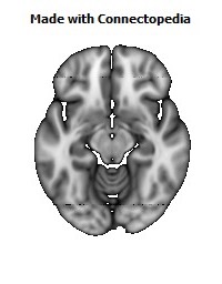
Structure
The two occipital lobes are the smallest of four paired lobes in the human cerebral cortex. Located in the rearmost portion of the skull, the occipital lobes are part of the forebrain. The cortical lobes are not defined by any internal structural features, but rather by the bones of the head bone that overlie them. Thus, the occipital lobe is defined as the part of the cerebral cortex that lies underneath the occipital bone. (See the human brain article for more information.)
The lobes rest on the tentorium cerebelli, a process of dura mater that separates the cerebrum from the cerebellum. They are structurally isolated in their respective cerebral hemispheres by the separation of the cerebral fissure. At the front edge of the occipital are several lateral occipital gyri, which are separated by lateral occipital sulcus.
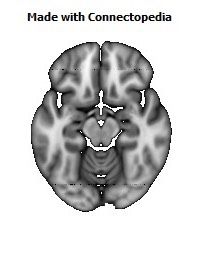
The occipital aspects along the inside face of each hemisphere are divided by the calcarine sulcus. Above the medial, Y-shaped sulcus lies the cuneus, and the area below the sulcus is the lingual gyrus.
Damage to the primary visual areas of the occipital lobe can leave a person with partial or complete blindness.
Function
The occipital lobe is divided into several functional visual areas. Each visual area contains a full map of the visual world. Although there are no anatomical markers distinguishing these areas (except for the prominent striations in the striate cortex), physiologists have used electrode recordings to divide the cortex into different functional regions.
The first functional area is the primary visual cortex. It contains a low-level description of the local orientation, spatial-frequency and color properties within small receptive fields. Primary visual cortex projects to the occipital areas of the ventral stream (visual area V2 and visual area V4), and the occipital areas of the dorsal stream—visual area V3, visual area MT (V5), and the dorsomedial area (DM).
A significant functional aspect of the occipital lobe is that it contains the primary visual cortex.
Retinal sensors convey stimuli through the optic tracts to the lateral geniculate bodies, where optic radiations continue to the visual cortex. Each visual cortex receives raw sensory information from the outside half of the retina on the same side of the head and from the inside half of the retina on the other side of the head. The cuneus (Brodmann's area 17) receives visual information from the contralateral superior retina representing the inferior visual field. The lingula receives information from the contralateral inferior retina representing the superior visual field. The retinal inputs pass through a "way station" in the lateral geniculate nucleus of the thalamus before projecting to the cortex. Cells on the posterior aspect of the occipital lobes' gray matter are arranged as a spatial map of the retinal field. Functional neuroimaging reveals similar patterns of response in cortical tissue of the lobes when the retinal fields are exposed to a strong pattern.
Clinical significance
If one occipital lobe is damaged, the result can be homonymous hemianopsia vision loss from similarly positioned "field cuts" in each eye. Occipital lesions can cause visual hallucinations. Lesions in the parietal-temporal-occipital association area are associated with color agnosia, movement agnosia, and agraphia. Damage to the primary visual cortex, which is located on the surface of the posterior occipital lobe, can cause blindness due to the holes in the visual map on the surface of the visual cortex that resulted from the lesions.
Epilepsy
Recent studies have shown that specific neurological findings have had an impact on idiopathic occipital lobe epilepsies. Occipital lobe seizures are triggered by a flash, or a visual image that contains multiple colors. These are called flicker stimulation (usually through TV) these seizures are referred to as photo-sensitivity seizures. Patients having experienced occipital seizures described their seizure as seeing bright colors, and having severe blurred vision (vomiting was also apparent in some patients). Occipital seizure are triggered mainly during the day, through television, video games or any flicker stimulatory system. Occipital seizures originate from an epileptic focus confined within the occipital lobes. They may be spontaneous or triggered by external visual stimuli. Occipital lobe epilepsies are etiologically idiopathic, symptomatic, or cryptogenic. Symptomatic occipital seizures can start at any age, as well as any stage after or during the course of the underlying causative disorder. Idiopathic occipital epilepsy usually starts in childhood. Occipital epilepsies account for approximately 5% to 10% of all epilepsies.