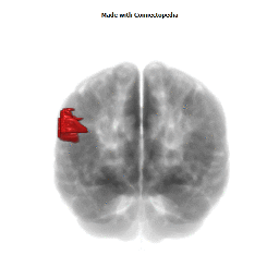

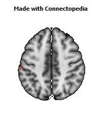
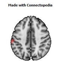
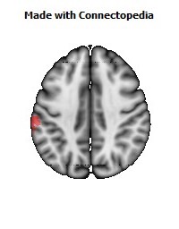
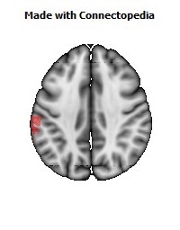
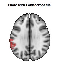
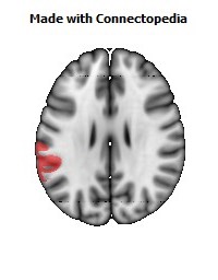
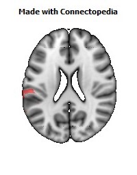

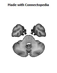
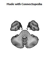
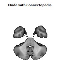
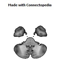


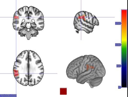
The supramarginal gyrus is a portion of the parietal lobe. It is probably involved with language perception and processing, and lesions in it may cause receptive aphasia.
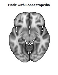
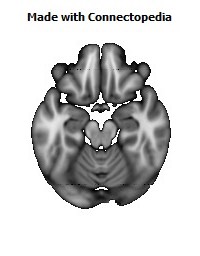
Parietal lobe
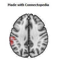
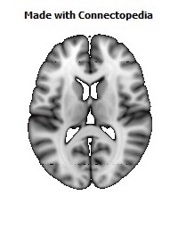
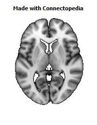
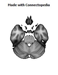
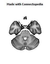
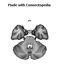
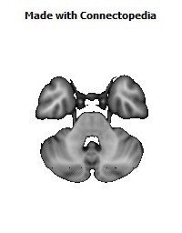
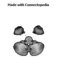
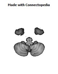
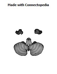
The parietal lobe is one of the four major lobes of the cerebral cortex in the brain of mammals. The parietal lobe is positioned above (superior to) the occipital lobe and behind (posterior to) the frontal lobe and central sulcus.
The parietal lobe integrates sensory information among various modalities, including spatial sense and navigation (See proprioception), the main sensory receptive area for the sense of touch (See somatosensation) in the somatosensory cortex which is just posterior to the central sulcus in the postcentral gyrus, and the dorsal stream of the visual system. The major sensory inputs from the skin (touch, temperature, and pain receptors) relay through the thalamus to parietal lobe.
Several portions of the parietal lobe are important in language processing. The somatosensory cortex can be illustrated as a distorted figure — the homunculus (Latin: "little man"), in which the body parts are rendered according to how much of the somatosensory cortex is devoted to them. The superior parietal lobule and inferior parietal lobule are the primary areas of body or spatial awareness. A lesion in the superior or inferior parietal lobule leads to hemineglect.
The name comes from the overlying parietal bone, which is named from the Latin paries-, "wall".
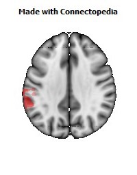
The parietal lobe is defined by three anatomical boundaries: The central sulcus separates the parietal lobe from the frontal lobe; the parieto-occipital sulcus separates the parietal and occipital lobes; the lateral sulcus (sylvian fissure) is the most lateral boundary, separating it from the temporal lobe; and the medial longitudinal fissure divides the two hemispheres. Within each hemisphere, the somatosensory cortex represents the skin area on the contralateral surface of the body.
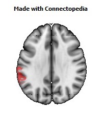
Immediately posterior to the central sulcus, and the most anterior part of the parietal lobe, is the postcentral gyrus (Brodmann area 3), the primary somatosensory cortical area. Dividing this and the posterior parietal cortex is the postcentral sulcus.
The posterior parietal cortex can be subdivided into the superior parietal lobule (Brodmann areas 5 + 7) and the inferior parietal lobule (39 + 40), separated by the intraparietal sulcus (IPS). The intraparietal sulcus and adjacent gyri are essential in guidance of limb and eye movement, and—based on cytoarchitectural and functional differences—is further divided into medial (MIP), lateral (LIP), ventral (VIP), and anterior (AIP) areas.
Function
The parietal lobe plays important roles in integrating sensory information from various parts of the body, knowledge of numbers and their relations, and in the manipulation of objects. Its function also includes processing information relating to the sense of touch. Portions of the parietal lobe are involved with visuospatial processing. Although multisensory in nature, the posterior parietal cortex is often referred to by vision scientists as the dorsal stream of vision (as opposed to the ventral stream in the temporal lobe). This dorsal stream has been called both the "where" stream (as in spatial vision) and the "how" stream (as in vision for action). The posterior parietal cortex (PPC) receives somatosensory and/or visual input, which then, through motor signals, controls movement of the arm, hand, as well as eye movements.
Various studies in the 1990s found that different regions of the posterior parietal cortex in Macaques represent different parts of space.
• The lateral intraparietal (LIP) contains a map of neurons (retinotopically-coded when the eyes are fixed) representing the saliency of spatial locations, and attention to these spatial locations. It can be used by the oculomotor system for targeting eye movements, when appropriate.
• The ventral intraparietal (VIP) area receives input from a number of senses (visual, somatosensory, auditory, and vestibular). Neurons with tactile receptive fields represent space in a head-centered reference frame. The cells with visual receptive fields also fire with head-centered reference frames but possibly also with eye-centered coordinates
• The medial intraparietal (MIP) area neurons encode the location of a reach target in nose-centered coordinates.
• The anterior intraparietal (AIP) area contains neurons responsive to shape, size, and orientation of objects to be grasped as well as for manipulation of hands themselves, both to viewed and remembered stimuli. The AIP has neurons that are responsible for grasping and manipulating objects through motor and visual inputs. The AIP and ventral premotor working together, are responsible for visuomotor transformations for actions of the hand.
More recent FMRI studies have shown that humans have similar functional regions in and around the intraparietal sulcus and parietal-occipital junction. The human "parietal eye fields" and "parietal reach region", equivalent to LIP and MIP in the monkey, also appear to be organized in gaze-centered coordinates so that their goal-related activity is "remapped" when the eyes move. Both the left and right parietal systems play a determining role in self transcendence, the personality trait measuring predisposition to spirituality.
This lobe is divided into two hemispheres—left and right. The left hemisphere is involved in symbolic functions in language and mathematics. Meanwhile, the right hemisphere is specialised to carry out images and understanding of maps (i.e. spatial relationships).
Clinical significance
Damage to the right hemisphere of this lobe results in the loss of imagery, visualization of spatial relationships and neglect of left-side space and left side of the body. Even drawings may be neglected on the left side. Damage to the left hemisphere of this lobe will result in problems in mathematics, long reading, writing, and understanding symbols. The parietal association cortex enables individuals to read, write, and solve mathematical problems.The sensory inputs from the right side of the body go to the left side of the brain and vice-versa.
Gerstmann's syndrome is associated with lesion to the dominant (usually left) parietal lobe. Balint's syndrome is associated with bilateral lesions. The syndrome of hemispatial neglect is usually associated with large deficits of attention of the non-dominant hemisphere. Optic ataxia is associated with difficulties reaching toward objects in the visual field opposite to the side of the parietal damage. Some aspects of optic ataxia have been explained in terms of the functional organization described above.
Apraxia is a disorder of motor control which can be referred neither to “elemental” motor deficits nor to general cognitive impairment. The concept of apraxia was shaped by Hugo Liepmann about a hundred years ago. Apraxia is predominantly a symptom of left brain damage, but some symptoms of apraxia can also occur after right brain damage.
Amorphosynthesis is a loss of perception on one side of the body caused by a lesion in the parietal lobe. Usually, left-sided lesions cause agnosia, a full-body loss of perception, while right-sided lesions cause lack of recognition of the person’s left side and extrapersonal space. The term amorphosynthesis was coined by D. Denny-Brown to describe patients he studied in the 1950s.
Can also result in sensory impairment when one of your senses (sight, hearing, smell, touch, taste and spatial awareness) is no longer normal.
Several studies have suggested that abnormal parietal function may be associated with schizophrenia. There is a possibility that grey matter abnormalities begin in parietal and occipital lobes, progress towards the frontal regions, causing schizophrenia structural and functional alterations.
Receptive aphasia
Receptive aphasia, also known as Wernicke’s aphasia, fluent aphasia, or sensory aphasia, is a type of aphasia traditionally associated with neurological damage to Wernicke’s area in the brain, (Brodmann area 22, in the posterior part of the superior temporal gyrus of the dominant hemisphere). However, the key deficits of receptive aphasia do not come from damage to Wernicke's area; instead, most of the core difficulties are proposed to come from damage to the medial temporal lobe and underlying white matter. Damage in this area not only destroys local language regions but also cuts off most of the occipital, temporal, and parietal regions from the core language region.
People with receptive aphasia are unable to understand language in its written or spoken form, and even though they can speak with normal grammar, syntax, rate, and intonation, they cannot express themselves meaningfully using language.
Receptive aphasia is not to be confused with Wernicke-Korsakoff syndrome.
Presentation
When we want to speak, we formulate what we are going to say in Wernicke’s area, which then transmits our plan of speech to Broca’s area, where the plan of speech is carried out. Wernicke’s Area is located posterior to the lateral sulcus, typically in the left hemisphere, between the visual, auditory, and somesthetic areas of the cerebral cortex. A person with this aphasia speaks normally but uses random or invented words; leaves out key words; substitutes words or verb tenses, pronouns, or prepositions; and utters sentences that do not make sense. They have normal sentence length and intonation but without true meaning. They can also have a tendency to talk excessively. A person with this aphasia cannot understand the spoken words of others or read written words. Speech is preserved, but language content is incorrect. Substitutions of one word for another (paraphasias, e.g. “telephone” for “television”) are common. Comprehension and repetition are poor.
Patients who recover from Wernicke’s aphasia report that, while aphasic, they found the speech of others to be unintelligible. And, despite being cognizant of the fact that they were speaking, they could neither stop themselves nor understand their own words.
The ability to understand and repeat songs is usually unaffected, as these are processed by the opposite hemisphere.
Patients also generally have no trouble purposefully reciting anything they have memorized. The ability to utter profanity is also left unaffected, however the patient typically has no control over it, and may not even understand their own profanity.
Damage to the posterior portion of the left hemisphere’s superior and middle temporal lobe or gyrus and the temporoparietal cortex can produce a lesion to Wernicke’s area and may cause fluent aphasia, or Wernicke’s aphasia. If Wernicke’s area is damaged in the non-dominant hemisphere, the syndrome resulting will be sensory dysprosody— the inability to perceive the pitch, rhythm, and emotional tone of speech.
Patients who communicated using sign language before the onset of the aphasia experience analogous symptoms.
The symptoms of Wernicke’s Aphasia reveal how important language is because people with the aphasia cannot express their thoughts. Some patients with the disorder do find a way to overcome this road block, and use facial expression and motor gestures to communicate instead.
Luria's theory
Luria proposed that this type of aphasia has three characteristics.
• 1) A deficit in the categorization of sounds. In order to hear and understand what is said, one must be able to recognize the different sounds of spoken language. For example, hearing the difference between bad and bed is easy for native English speakers. The Dutch language however, makes a much greater difference in pronunciation between these vowels, and therefore the Dutch have difficulties hearing the difference between them in English pronunciation. This problem is exactly what patients with Wernicke’s aphasia have in their own language: they can't isolate significant sound characteristics and classify them into known meaningful systems.
• 2) A defect in speech. A patient with Wernicke's aphasia can and may speak a great deal, but he or she confuses sound characteristics, producing “word salad” in extreme cases: intelligible words that appear to be strung together randomly.
• 3) An impairment in writing. A person who cannot discern sounds cannot be expected to write.