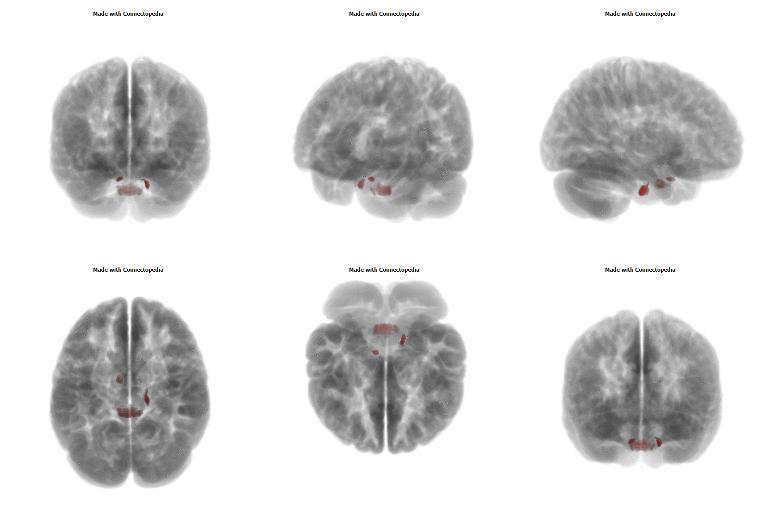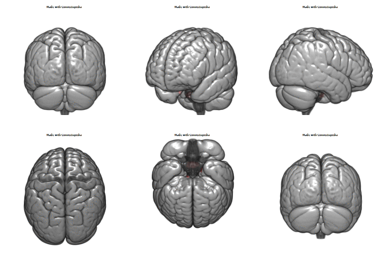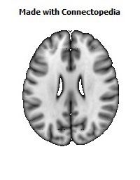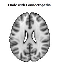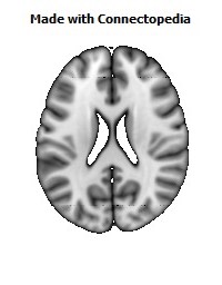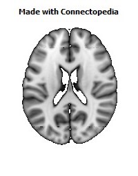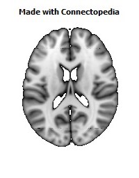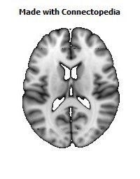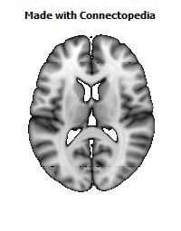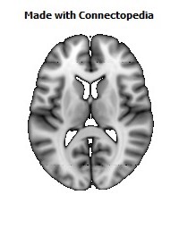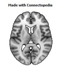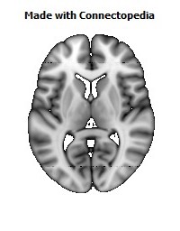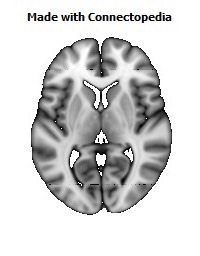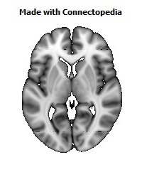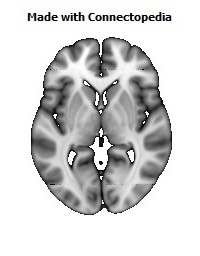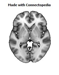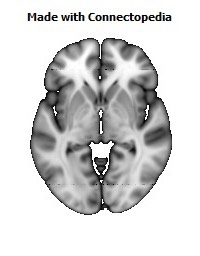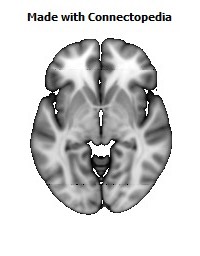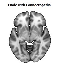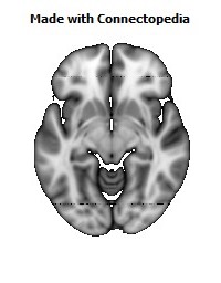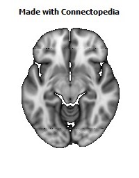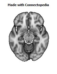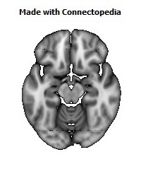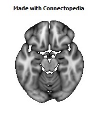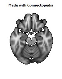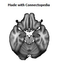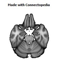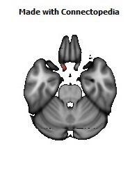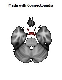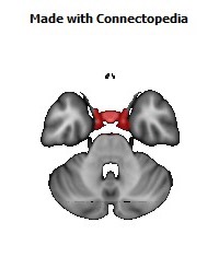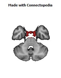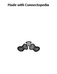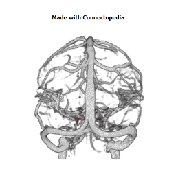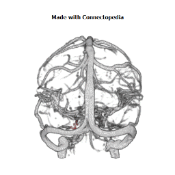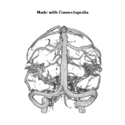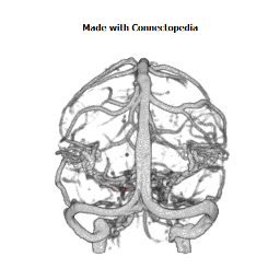
The cavernous sinus (or lateral sellar compartment), within the human head, is a large collection of thin-walled veins creating a cavity bordered by the temporal bone of the skull and the sphenoid bone, lateral to the sella turcica.
Structure
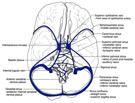
The sinus may be joined by several anastomoses across the midline. The cavernous sinus receives blood via the superior and inferior ophthalmic veins through the superior orbital fissure and from superficial cortical veins, and is connected to the basilar plexus of veins posteriorly. The internal carotid artery (carotid siphon), and cranial nerves III, IV, V (branches V1 and V2) and VI all pass through this blood filled space. Infection from the face may reach the cavernous sinus through its many anastomotic connections, with severe consequences. The cavernous sinus drains by two larger channels, the superior and inferior petrosal sinuses, ultimately into the internal jugular vein via the sigmoid sinus. Also drains with emissary vein to pterygoid plexus.
Function
Each cavernous sinus (one for each hemisphere of the brain) contains the following:
- vertically, from superior to inferior (within the lateral wall of the sinus)
- oculomotor nerve (CN III)
- trochlear nerve (CN IV)
- ophthalmic nerve, the V1 branch of the trigeminal nerve (CN V)
- maxillary nerve, the V2 branch of CN V
Unlike the nerves listed above, the abducens nerve (CN VI) does not run within the lateral wall of the cavernous sinus; rather, it runs through the middle of the sinus alongside the internal carotid artery.
These nerves, with the exception of CN V2, pass through the cavernous sinus to enter the orbital apex through the superior orbital fissure. The maxillary nerve, division V2 of the trigeminal nerve travels through the lower portion of the sinus and exits via the foramen rotundum.
- horizontally, from medial to lateral
- internal carotid artery (and sympathetic plexus). See also cavernous part of internal carotid artery.
The optic nerve lies just above and outside the cavernous sinus, superior and lateral to the pituitary gland on each side, and enters the orbital apex via the optic canal.
Venous connections
It receives tributaries from:
- Superior and inferior ophthalmic veins
- Sphenoparietal sinus
- Superficial middle cerebral veins
The veins of exit are to the superior and inferior petrosal sinuses as well as via the emissary veins through the foramina of the skull (mostly through foramen ovale). There are also connections with the pterygoid plexus of veins via inferior ophthalmic vein, deep facial vein and emissary veins.
Clinical significance
It is the only anatomic location in the body in which an artery travels completely through a venous structure. If the internal carotid artery ruptures within the cavernous sinus, anarteriovenous fistula is created (more specifically, a carotid-cavernous fistula). Lesions affecting the cavernous sinus may affect isolated nerves or all the nerves traversing through it.
The pituitary gland lies between the two paired cavernous sinuses. An abnormally growing pituitary adenoma, sitting on the bony sella turcica, will expand in the direction of least resistance and eventually compress the cavernous sinus. Cavernous sinus syndrome may result from mass effect of these tumors and cause ophthalmoplegia (from compression of the oculomotor nerve, trochlear nerve, and abducens nerve), ophthalmic sensory loss (from compression of the ophthalmic nerve), and maxillary sensory loss (from compression of the maxillary nerve). A complete lesion of the cavernous sinus disrupts CN III, IV, and VI, causing total ophthalmoplegia, usually accompanied by a fixed, dilated pupil. Involvement of CN V (V1 and variable involvement of V2) causes sensory loss in these divisions of the trigeminal nerve. Horner's syndrome can also occur due to involvement of the carotid ocular sympathetics, but may be difficult to appreciate in the setting of a complete third nerve injury.
Because of its connections with the facial vein via the superior ophthalmic vein, it is possible to get infections in the cavernous sinus from an external facial injury within the Danger area of the face. In patients with thrombophlebitis of the facial vein, pieces of the clot may break off and enter the cavernous sinus, forming a cavernous sinus thrombosis. From there the infection may spread to the dural venous sinuses. Infections may also be introduced by facial lacerations and by bursting pimples in the areas drained by the facial vein.
Potential causes of cavernous sinus syndrome include metastatic tumors, direct extension of nasopharyngeal tumors, meningioma, pituitary tumors or pituitary apoplexy, aneurysms of the intracavernous carotid artery, cavernous-carotid arteriovenous fistula, bacterial infection causing cavernous sinus thrombosis, aseptic thrombosis, idiopathic granulomatous disease (Tolosa-Hunt syndrome), and fungal infections. Cavernous sinus syndrome is a medical emergency, requiring prompt medical attention, diagnosis, and treatment.
