
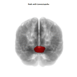


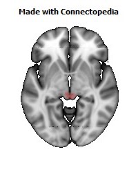
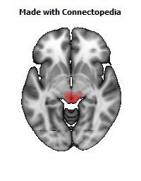
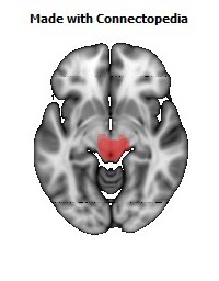
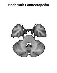
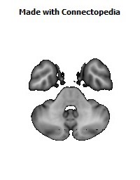
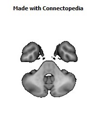
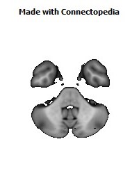
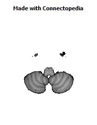


The great cerebral vein is one of the large blood vessels in the skull draining the cerebrum (brain). It is also known as the "vein of Galen" (VG), named for its discoverer, the Greek physician Galen. However, it is not the only vein with this eponym.
Structure
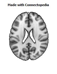
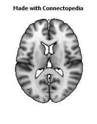
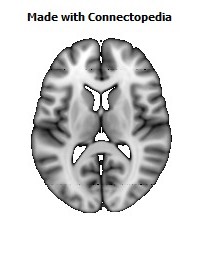
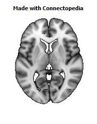
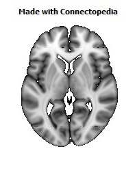
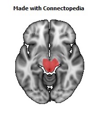
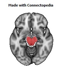
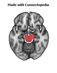
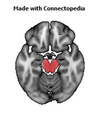
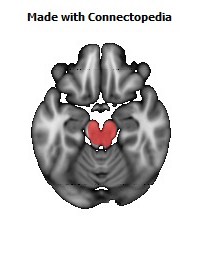
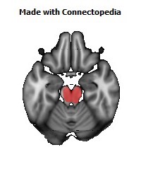
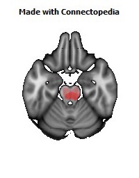
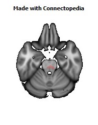
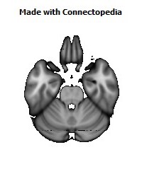
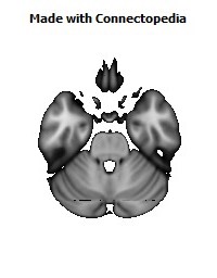
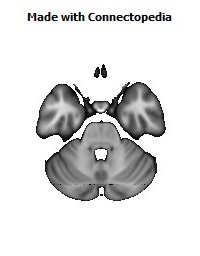
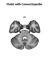
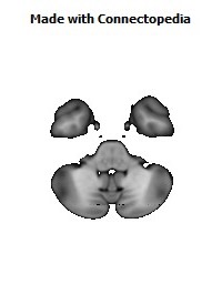
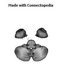
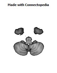
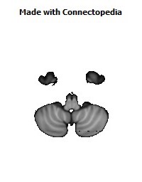
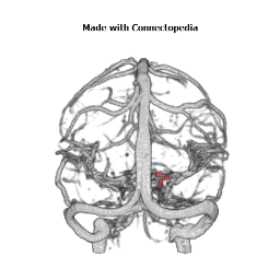
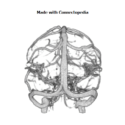
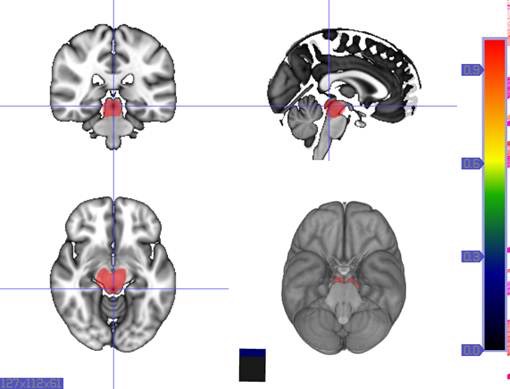
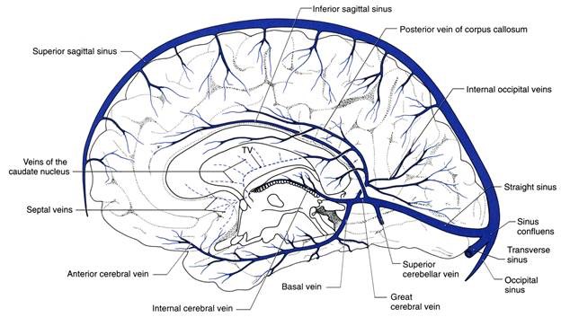
The great cerebral vein of Galen is considered a deep/internal cerebral vein. The internal cerebral veins are formed by the union of the thalamostriate vein and the choroid vein at the interventricular foramen. The deep/internal group of cerebral veins can be seen on the superior surfaces of the caudate nuclei and thalami just under the corpus callosum. The veins at the anterior poles of the thalami merge posterior to the pineal gland to form the Great Cerebral Vein of Galen. Most of the blood in the deep cerebral veins collects into the great cerebral vein. This comes from the inferior side of the posterior end of the corpus callosum and empties into the straight sinus located in the midline of the tentorium. Unlike the arteries, the cerebral veins have anastomoses. With extensive anastomoses, a slow occlusion of a venous channel rarely produces more than transitory effects.
There are both superficial/external and deep/internal cerebral veins, the great cerebral vein being a deep/internal vein as mentioned above) in the brain. As there are similarities, there are also differences between these two types of veins in the brain. The superficial veins at the dorsal parts of the hemispheres run upward and medially and empty into the large superior sagittal sinus in the upper margin of the falx cerebri. The superior sagittal sinus divides into two parts called the transverse sinuses where the falx cerebri meets the tentorium cerebelli. The sigmoid sinus, which continues the transverse sinus, empties into the jugular vein at the jugular foramen. The internal jugular vein leaves the skull and travels downward to the neck.
The veins of the brain have very thin walls and contain no valves. They emerge in the brain and lie in the subarachnoid space. They pierce the arachnoid matter and the meningeal layer in the dura and drain into the cranial venous sinuses.
Clinical relevance
Malformations
Most conditions associated with the great cerebral vein are due to congenital defects. Vein of Galen aneurysmal malformations (VGAM) are the most common form of symptomatic cerebrovascular malformation in neonates and infants. The presence and locations of angiomas are very variable and do not follow any predictable pattern. The congenital malformation develops during weeks 6-11 of fetal development as a persistent embryonic prosencephalic vein of Markowski; thus, VGAM is actually a misnomer. The vein of Markowski actually drains into the vein of Galen.
Absence
Absence of the great cerebral vein is a type of malformation. The veins of the deep structures of the brain normally drain through the Galenic system. In the absence of the great cerebral vein, the veins from the diencephalon and the basal ganglia drain laterally into the transverse sinus instead of conjoining in the midline through the Galenic drainage system. Absence of the great cerebral vein is quite rare. It is detected in infancy. Most patients die in the neonatal period or in early infancy.
Thrombosis
Thrombosis of the great cerebral vein is a form of stroke due to a blood clot in the vein. It affects just 3 to 8% of patients, predominantly women. Patients may present with consciousness problems, headaches, nausea, visual defects, fatigue, disturbance of eye movements and pupillary reflexes, or coma. Thrombosis of the cerebral vein is often deadly but can be survived. Risk factors include oral contraceptives, pregnancy, and the puerperium.