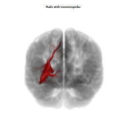
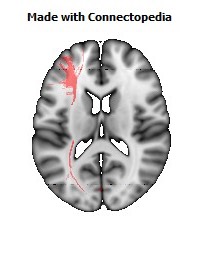
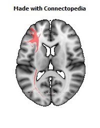
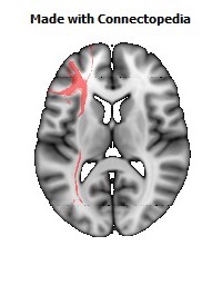
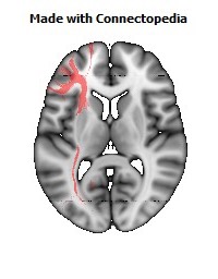
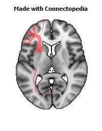
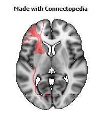
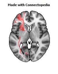
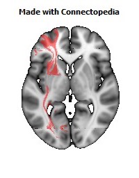
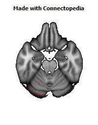


The occipitofrontal fasciculus passes backward from the frontal lobe, along the lateral border of the caudate nucleus, and on the medial aspect of the corona radiata; its fibers radiate in a fan-like manner and pass into the occipital and temporal lobes lateral to the posterior and inferior cornua.
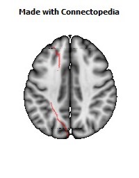
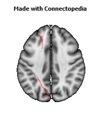
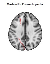
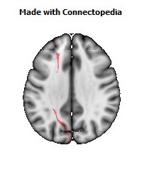
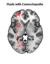
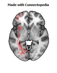
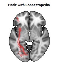
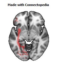
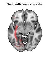
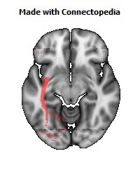
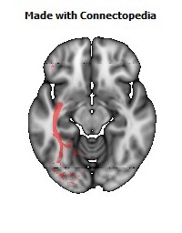
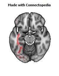
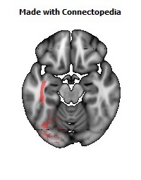
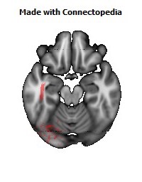
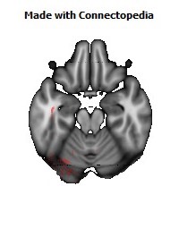
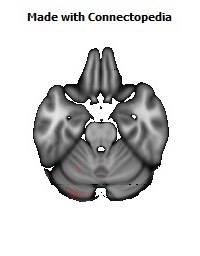
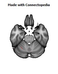
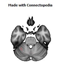
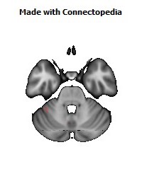
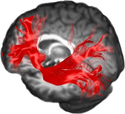
The inferior longitudinal fasciculus (ILF) was classically defined as a direct connection from the occipital cortex to the temporal lobe. But even after the advent of diffusion tensor imaging (DTI) this view was challenged by several authors. Using DTI and tractography, which allows for a non-invasive assessment of the microstructural organization of white matter pathways, Catani et al. elegantly confirmed the direct pathway between the occipital cortex and temporal lobe in a group average, as well as in eleven individual normal controls. The ILF fiber bundle connects the occipital lobe with the anterior part of the temporal lobe, running laterally and inferiorly above optic radiation fibers. However, in addition to the ILF there is a second major fiber bundle connecting the occipital cortex to the frontal brain known as the inferior fronto-occipital fasciculus (IFOF) that runs medially and above the optic pathways and spatially overlaps with the ILF along part of their pathways. The IFOF is known as a direct pathway, one that connects the occipital, posterior temporal, and the orbito-frontal areas. The ILF, on the other hand, is considered to be an indirect pathway essentially connecting similar brain areas and anteriorly joins the uncinate fasciculus to relay information to the orbito-frontal brain. In a recent report, Wahl et al. studied whether specific patterns of correlation and overlap among the major brain fibers bundles exist, using DTI scalar parameters such as fractional anisotropy, and axial, radial, and mean diffusivity. The only significant overlaps they found were between the IFOF and the ILF, and between the IFOF and the uncinate fasciculus. Such overlap may indeed reflect functional similarity as well as spatially sharing overlapping paths.
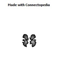
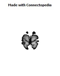
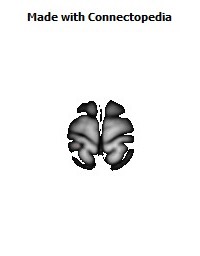
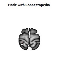
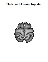
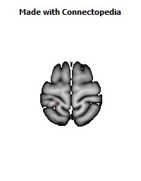
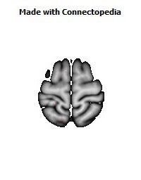
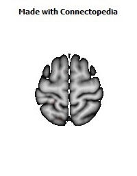
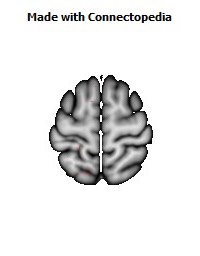
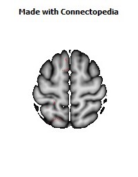
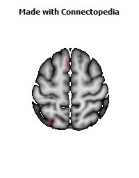
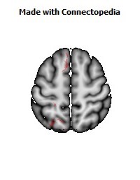
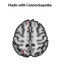
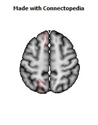
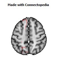
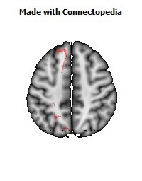
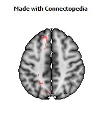
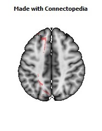
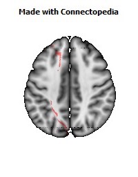
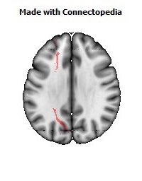
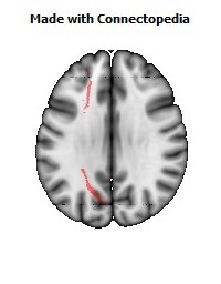
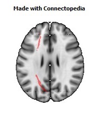
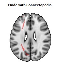
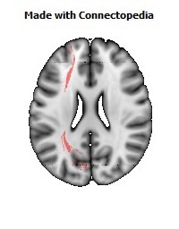
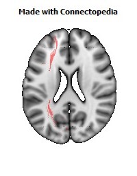
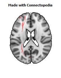
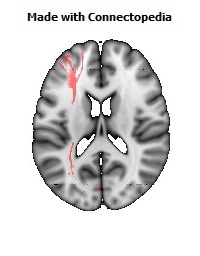
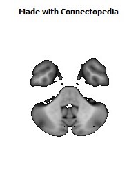
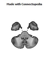
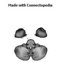
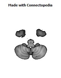
Both Left and Right fasciculi are asymmetric:
