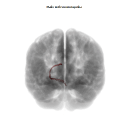


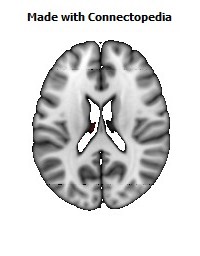
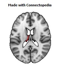
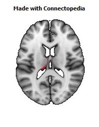
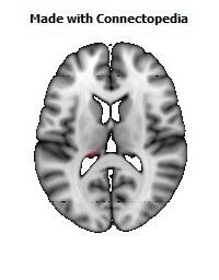
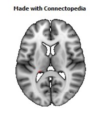
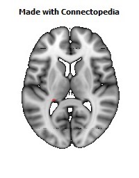
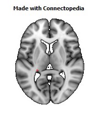
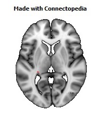
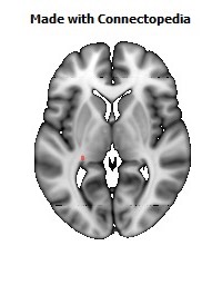
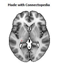
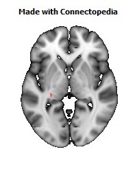
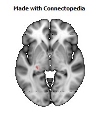
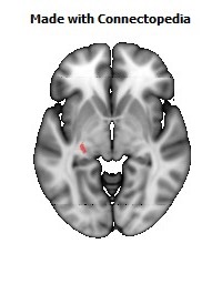
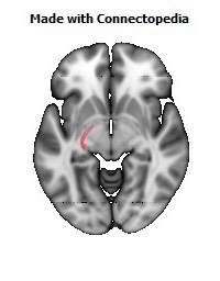
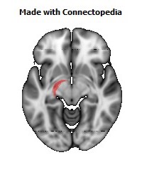
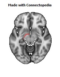
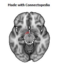
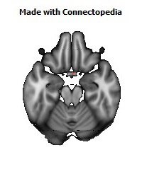

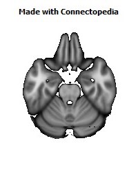
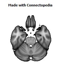
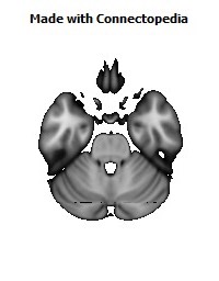
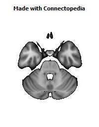
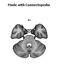
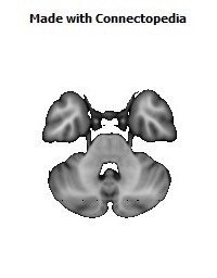
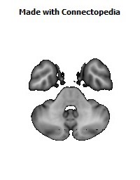
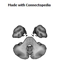
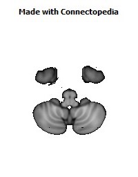
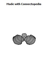
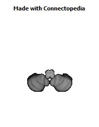


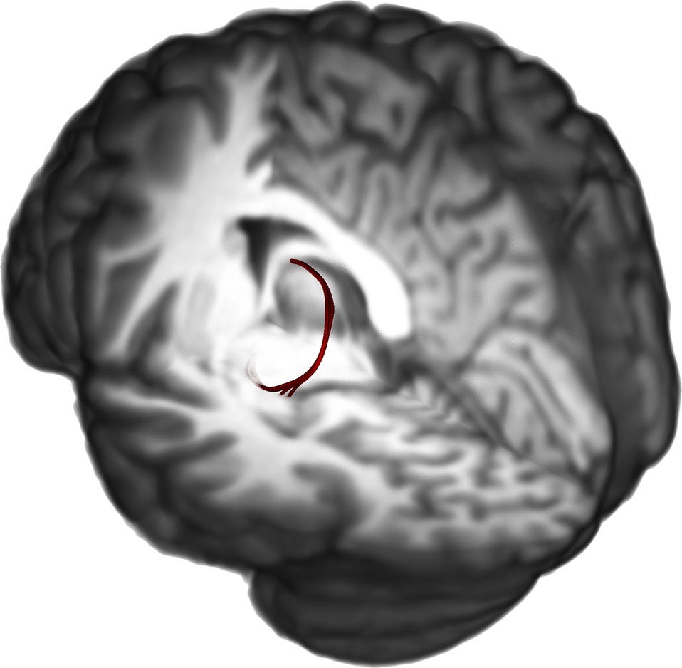
The stria terminalis (or terminal stria) is a structure in the brain consisting of a band of fibers running along the lateral margin of the ventricular surface of the thalamus. Serving as a major output pathway of the amygdala, the stria terminalis runs from its centromedial division to the ventral medial nucleus of the hypothalamus.
Anatomy
The stria terminalis covers the thalamostriate vein, marking a line of separation between the thalamus and the caudate nucleus as seen upon gross dissection of the ventricles of the brain, viewed from the superior aspect.
The stria terminalis extends from the region of the interventricular foramen to the temporal horn of the lateral ventricle, carrying fibers from the amygdala to the septal nuclei, hypothalamic, and thalamic areas of the brain. It also carries fibers projecting from these areas back to the amygdala.
Functions
The activity of the bed nucleus of the stria terminalis correlates with anxiety in response to threat monitoring. It is thought to act as a relay site within the hypothalamic-pituitary-adrenal axis and regulate its activity in response to acute stress. It is also thought to promote behavioral inhibition in response to unfamiliar individuals, by input from the orbitofrontal cortex. Bilateral disruption of this pathway has been shown to attenuate reinstatement of drug seeking behaviour in rodents.
Bed nucleus of the stria terminalis (BNST)
The central subdivision of the bed nucleus of the stria terminalis (BSTc) is sexually dimorphic. On average, the BSTc is twice as large in men as in women and contains twice the number of somatostatin neurons. A sample of six male-to-female transsexuals taking estrogen were found to have female-typical number of cells in the BSTc, whereas a female-to-male transsexual taking testosterone was found to have a male-typical number. The authors (W. Chung, G. De Vries, Dick Swaab) also examined subjects with hormone-related disorders and found no pattern between those disorders and the BSTc while the single untreated male-to-female transsexual had a female-typical number of cells. They concluded that the BSTc provides evidence for a neurobiological basis of gender identity disorder and proposed that such was determined before birth.
Criticisms of the former statement suggest the study used an unrepresentative sample and did not adequately control for hormone replacement therapy, which has been shown to influence hypothalamic size, even though the study tried to do this by including non-transsexual male and female controls which, for a variety of medical reasons, had experienced hormone reversal. The statement about the neurobiological basis from birth has later been brought to question, though not refuted, by a follow up study by the same group which found that the sexual dimorphism of the BSTc is not present before adulthood (approximately 22 years of age) even though transsexuals report being aware of their gender identity since childhood.
Reduction of the size of the bed nucleus of the stria terminalis has been observed in pedophilic male perpetrators, in addition to reductions in the right amygdala, hypothalamus and abnormalities in related structures. The authors propose that childhood deficits in the BNST and medial amygdala may cause inhibition of sexual maturity.