
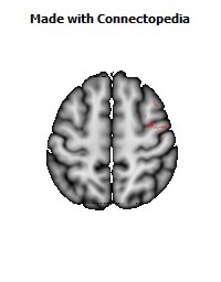
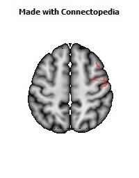
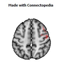
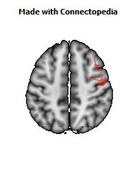
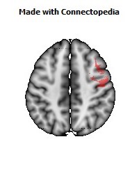
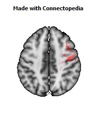
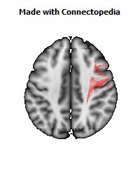
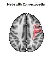
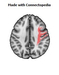
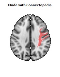
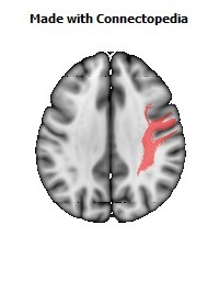
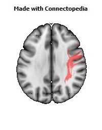
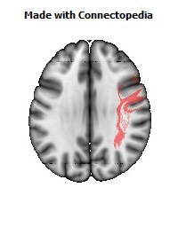
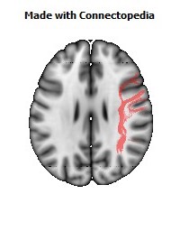
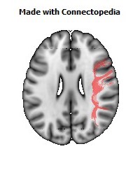
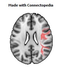
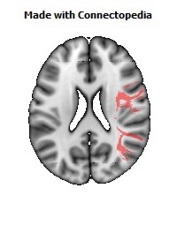
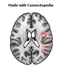
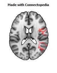
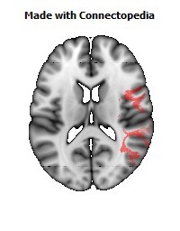
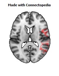
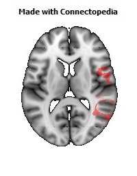
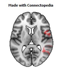
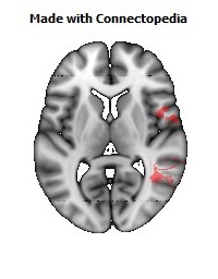
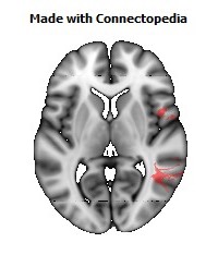
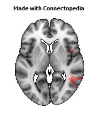
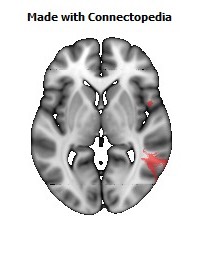
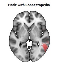
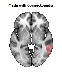
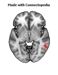
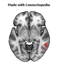
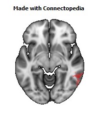
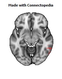
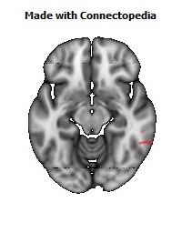
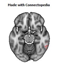
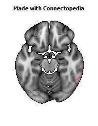

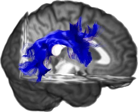
The superior longitudinal fasciculus (also called the superior longitudinal fascicle or SLF) is a pair of long bi-directional bundles of neurons connecting the front and the back of the cerebrum. Each association fiber bundle is lateral to the centrum ovale of a cerebral hemisphere and connects the frontal, occipital, parietal, and temporal lobes. The neurons pass from the frontal lobe through the operculum to the posterior end of the lateral sulcus where numerous neurons radiate into the occipital lobe and other neurons turn downward and forward around the putamen and radiate to anterior portions of the temporal lobe.
Anatomy
The SLF is composed of four distinct components SLF I, SLF II, SLF III, and arcuate fascicle (AF). In humans, these four components are bundled together although they are functionally separate. In non-human primates, the SLF and AF are anatomically separate and have separate trajectories.
SLF I
SLF I is the dorsal component and originates in the superior and medial parietal cortex, passes around the cingulate sulcus and in the superior parietal and frontal white matter, and terminates in the dorsal and medial cortex of the frontal lobe (Brodmann 6, 8, and 9) and in the supplementary motor cortex (M II).
SLF II
SLF II is the major component of SLF and originates in the caudal-inferior parietal cortex and terminates in the dorsolateral prefrontal cortex (Brodmann 6, 8 and 46).
SLF III
SLF III is the ventral component and originates in the supramarginal gyrus (rostral portion of the inferior parietal lobe) and terminates in the ventral premotor and prefrontal cortex (Brodmann 6, 44, and 46).
AF
The AF originates in the caudal area of the superior temporal gyrus and passes next to the neurons of SLF II above the Sylvian fissure and insula in non-human primates. In humans, neurons that originate from the caudal superior temporal gyrus and the superior temporal sulcus pass around the caudal Sylvian fissure and along with the SLF bundle and terminate in the dorsal prefrontal cortex (Brodmann areas 8 and 46).
Functions
SLF I
SLF I connects to the superior parietal cortex which encodes locations of body parts in a body-centric coordinate system and with M II and dorsal premotor cortex. This suggests the SLF I is involved with regulating motor behavior, especially conditional associative tasks which select among competing motor tasks based on conditional rules.
SLF II
SLF II connects to the caudal inferior parietal cortex which controls spatial attention and visual and oculomotor functions. This suggests the SLF II provides the prefrontal cortex with parietal cortex information regarding perception of visual space. Since these bundles are bi-directional, working memory (Brodmann 46) in the prefrontal cortex may provide the parietal cortex with information to focus spatial attention and regulate selection and retrieval of spatial information.
SLF III
SLF III connects the rostral inferior parietal cortex which receives information from the ventral precentral gyrus. This suggests that the SLF III transfers somatosensory information, such as language articulation, between the ventral premotor cortex, Brodmann 44 (pars opercularis), the supramarginal gyrus (Brodmann 40), and the laterial inferior prefrontal cortex working memory (Brodmann 46).
AF
The arcuate fascicle connects the superior temporal gyrus (Tpt) with the dorsal prefrontal cortex which suggests auditory information is transmitted between those two areas of cortex.
Both Left and Right fasciculi are asymmetric:
