


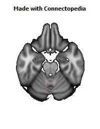
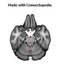
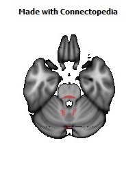
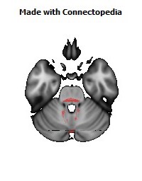
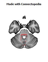
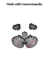
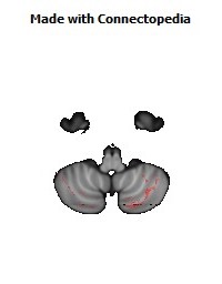
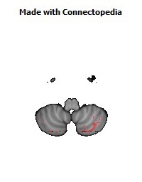

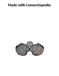

The pons /ˈpɒnz/ is part of the brainstem that links the medulla oblongata and the thalamus. It is also called the pons Varolii ("bridge of Varoli"), after the Italian anatomist and surgeon Costanzo Varolio (1543–75). It is cranial to the medulla oblongata, caudal to the midbrain, and ventral to the cerebellum. In humans and other bipeds, this means it is above the medulla, below the midbrain, and anterior to the cerebellum. This white matter includes tracts that conduct signals from the cerebrum down to the cerebellum and medulla, and tracts that carry the sensory signals up into the thalamus.
The pons in humans measures about 2.5 cm or 1 inch in length. Most of it appears as a broad anterior bulge rostral to the medulla. Posteriorly, it consists mainly of two pairs of thick stalks called cerebellar peduncles. They connect the cerebellum to the pons and midbrain.
The pons contains nuclei that relay signals from the forebrain to the cerebellum, along with nuclei that deal primarily with sleep, respiration, swallowing, bladder control, hearing, equilibrium, taste, eye movement, facial expressions, facial sensation, and posture.
Within the pons is the pneumotaxic center, a nucleus that regulates the change from inhalation to exhalation.
The pons is implicated in sleep paralysis, and also plays a role in generating dreams.
Structure
Development
During embryonic development, the embryonic metencephalon develops from the rhombencephalon and gives rise to two structures: the pons and the cerebellum. The alar plate produces sensory neuroblasts, which will give rise to the solitary nucleus and its special visceral afferent (SVA) column; the cochlear and vestibular nuclei, which form the special somatic afferent (SSA) fibers of the vestibulocochlear nerve, the spinal and principal trigeminal nerve nuclei, which form the general somatic afferent column (GSA) of the trigeminal nerve, and the pontine nuclei which relays to the cerebellum.
Basal plate neuroblasts give rise to the abducens nucleus,which forms the general somatic efferent fibers (GSE); the facial and motor trigeminal nuclei, which form the special visceral efferent (SVE) column, and the superior salivatory nucleus, which forms the general visceral efferent fibers of the facial nerve.
Nucleus
A number of cranial nerve nuclei are present in the pons:
• mid-pons: the 'chief' or 'pontine' nucleus of the trigeminal nerve sensory nucleus (V)
• mid-pons: the motor nucleus for the trigeminal nerve (V)
• lower down in the pons: abducens nucleus (VI)
• lower down in the pons: facial nerve nucleus (VII)
• lower down in the pons: vestibulocochlear nuclei (vestibular nuclei and cochlear nuclei) (VIII)
The pontine nuclei (or griseum pontis) are a part of the pons involved in motor activity. Corticopontine fibres carry information from the primary motor cortex to the ipsilateral pontine nucleus in the ventral pons, and the pontocerebellar projection then carries that information to the contralateral cerebellum via the middle cerebellar peduncle. Extension of these nuclei in the medulla oblongata are named arcuate nucleus (medulla) which has the same function.
They therefore allow modification of actions in the light of their outcome, or error correction, and are hence important in learning motor skills.
Function
The functions of these four nerves include sensory roles in hearing, equilibrium, and taste, and in facial sensations such as touch and pain, as well as motor roles in eye movement, facial expressions, chewing, swallowing, and the secretion of saliva and tears.
Clinical significance
• Central pontine myelinosis is a demyelination disease that causes difficulty with sense of balance, walking, sense of touch, swallowing and speaking. In a clinical setting, it is often associated with transplant. Undiagnosed, it can lead to death or locked-in syndrome.
Other animals
Evolution
The pons first evolved as an offshoot of the medullary reticular formation.[4] Since lampreys possess a pons, it has been argued that it must have evolved as a region distinct from the medulla by the time the first agnathans appeared, 505 million years ago.
Pontine tegmentum
The pontine tegmentum is a part of the pons of the brain involved in the initiation of REM sleep. It includes the pedunculopontine nucleus and the laterodorsal tegmental nucleus, among others, and is located near the raphe nucleus and the locus ceruleus.
The trapezoid body, involved in binaural hearing, is part of the pontine tegmentum.
Function
In animal studies, lesions of the pontine tegmentum greatly reduce or even eliminate REM sleep. Injection of a cholinergic agonist (e.g. carbachol), into the pontine tegmentum produces a state of REM sleep in cats.
PET studies seem to indicate that there is a correlation between blood flow in the pontine tegmentum, REM sleep, and dreaming.
Pontine waves (P-waves, or ponto-geniculate-occipital waves) are brain waves generated in the pontine tegmentum. They can be observed in mammals, precede the onset of REM sleep, and continue throughout its course. After periods of memory training, P-wave density increases during subsequent sleep periods in rats. This may be an indication of a link between sleep and learning.