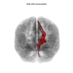

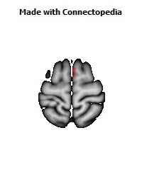
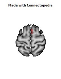
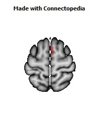
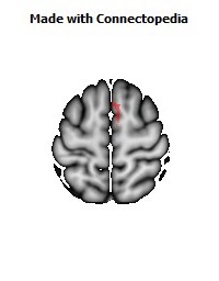
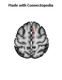
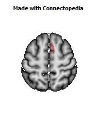
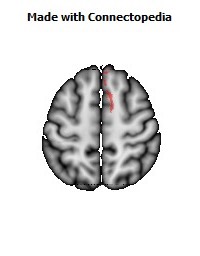
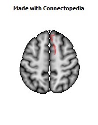
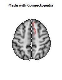
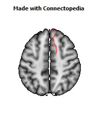
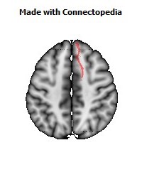
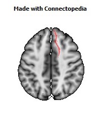
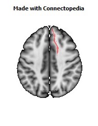
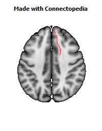
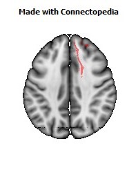
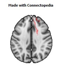
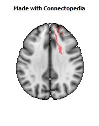
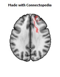

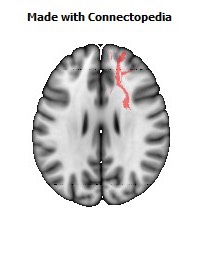
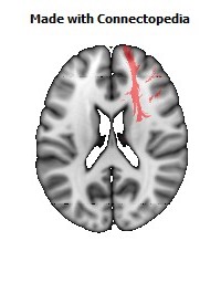
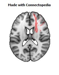
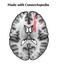
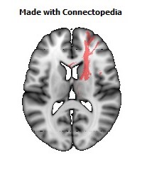
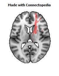
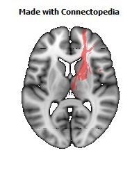
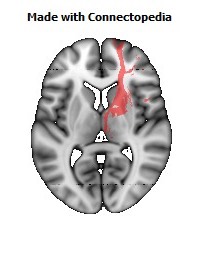
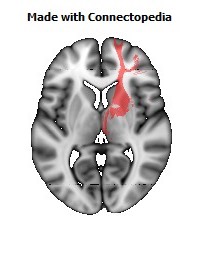
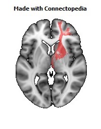
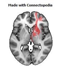
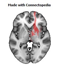
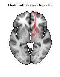
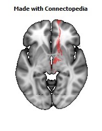
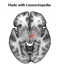
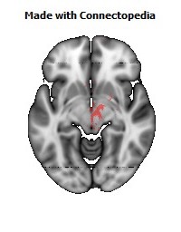
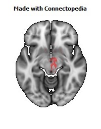
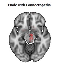
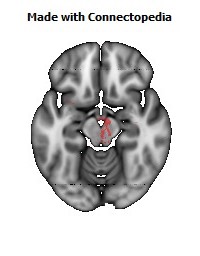
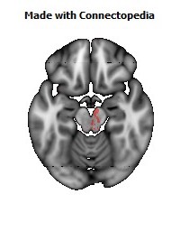
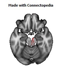
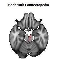
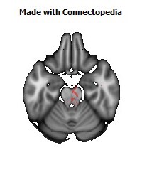
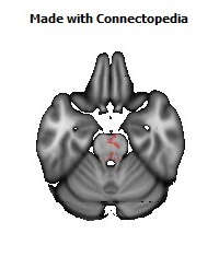
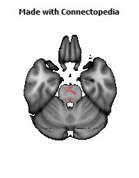
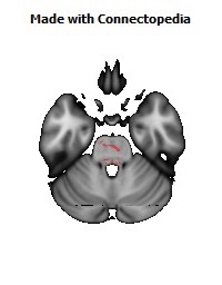
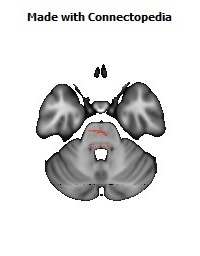
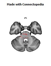
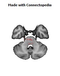
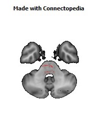
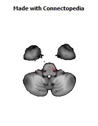

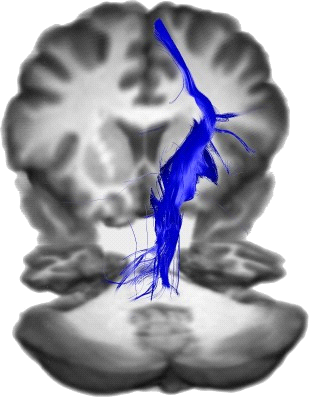
The internal capsule is a white matter structure in the brain that carries information past the basal ganglia, separating the caudate nucleus and the thalamus from the putamen and the globus pallidus. The internal capsule contains both ascending and descending axons.
The corticospinal tract constitutes a large part of the internal capsule, carrying motor information from the primary motor cortex to the lower motor neurons in the spinal cord. Above the basal ganglia the corticospinal tract is a part of the corona radiata, below the basal ganglia the tract is called crus cerebri (a part of the cerebral peduncle) and below pons it is referred to as the corticospinal tract.
Structure
The internal capsule is V-shaped when cut horizontally, in a transverse plane.
When cut horizontally:
• the bend in the V is called the genu
• the anterior limb or crus anterius is the part in front of the genu, between the head of the caudate nucleus and the lenticular nucleus
• the posterior limb or crus posterius is the part behind the genu, between the thalamus and lenticular nucleus
• the retrolenticular portion is caudal to the lenticular nucleus and carries optic tracts including the geniculocalcarine radiations.
• the sublenticular portion is beneath the lenticular nucleus and are tracts involved in the auditory pathway from medial geniculate nucleus to the primary auditory cortex (Brodmann Area 41)
Genu
The genu of internal capsule is the flexure of the internal capsule.
The fibers in the region of the genu are named the geniculate fibers; they originate in the motor part of the cerebral cortex, and, after passing downward through the base of the cerebral peduncle with the cerebrospinal fibers, undergo decussation and end in the motor nuclei of the cranial nerves of the opposite side.
It is formed by fibers from the corticonuclear tracts.
Anterior limb
The anterior limb of internal capsule (or frontal part) contains:
• (1) fibers running from the thalamus to the frontal lobe;
• (2) fibers connecting the lentiform and caudate nuclei;
• (3) fibers connecting the cortex with the corpus striatum; and
• (4) fibers passing from the frontal lobe through the medial fifth of the base of the cerebral peduncle to the nuclei pontis.
Posterior limb
The posterior limb of internal capsule (or occipital part) is the portion of the internal capsule posterior to the genu.
The anterior two-thirds of the occipital part of the internal capsule contains fibers of the corticospinal tract, which arise in the motor area of the cerebral cortex and, passing downward through the middle three-fifths of the base of the cerebral peduncle, are continued into the pyramids of the medulla oblongata.
The posterior third of the occipital part contains:
• (1) sensory fibers, largely derived from the thalamus, though some may be continued upward from the medial lemniscus;
• (2) the fibers of optic radiation, from the lower visual centers to the cortex of the occipital lobe;
• (3) acoustic fibers, from the lateral lemniscus to the temporal lobe; and
• (4) fibers which pass from the occipital and temporal lobes to the nuclei pontis.
Blood supply
The superior parts of both the anterior and posterior limbs and the genu of the internal capsule are supplied by the lenticulostriate arteries, which are branches of the M1 segment of the middle cerebral artery.
The inferior half of the anterior limb is supplied via the recurrent artery of Heubner, which is a branch of the anterior cerebral artery.
The inferior half of the posterior limb is supplied by the anterior choroidal artery, which is a branch of the internal carotid artery.
In summary, the blood supply of the internal capsule is
Anterior limb: lenticulostriate branches of middle cerebral artery (superior half) & recurrent artery of Heubner off of the anterior cerebral artery (inferior half) Genu: lenticulostriate branches of middle cerebral artery Posterior limb: lenticulostriate branches of middle cerebral artery (superior half) & anterior choroidal artery branch of the internal carotid artery (inferior half)
As in many parts of the body, some degree of variation in the blood supply exists. For example, thalamoperforator arteries, which are branches of the basilar artery, occasionally supply the inferior half of the posterior limb.
Function
Working anterior to posterior:
• The anterior limb of the internal capsule contains:
1) Frontopontine (corticofugal) fibers project from frontal cortex to pons;
2) Thalamocortical fibers (part of the thalamocortical radiations) connect the medial and anterior nuclei of the thalamus to the frontal lobes (these are severed during a prefrontal lobotomy).
• The genu contains corticobulbar fibers, which run between the cortex and the brainstem.
• The posterior limb of the internal capsule contains corticospinal fibers, sensory fibers (including the medial lemniscus and the anterolateral system) from the body and a few corticobulbar fibers.
Other fibers within the internal capsule
• The retrolenticular part contains fibers from the optic system, coming from the lateral geniculate nucleus of the thalamus. More posteriorly, this becomes the optic radiation. Some fibers from the medial geniculate nucleus (which carry auditory information) also pass in the retrolenticular internal capsule, but most are in the sublenticular part.
• The sublenticular part contains fibers connecting with the temporal lobe. These include the auditory radiations and temporopontine fibers.
Clinical significance
The lenticulostriate arteries supply a substantial amount of the internal capsule. These small vessels are particularly vulnerable to narrowing in the setting of chronic hypertension and can result in small, punctate infarctions or intraparenchymal haemorrhage due to vessel rupture.
Lesions of the genu of the internal capsule affect fibers of the corticobulbar tract.
The primary motor cortex sends its axons through the posterior limb of the internal capsule. Lesions, therefore, result in a contralateral hemiparesis or hemiplegia. While symptoms of weakness due to an isolated lesion of the posterior limb can initially be severe, recovery of motor function is sometimes possible due to spinal projections of premotor cortical regions that are contained more rostrally in the internal capsule.