

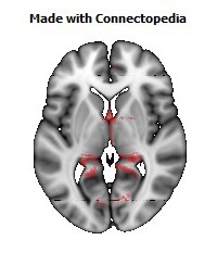
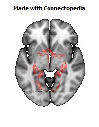
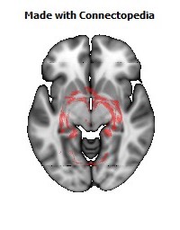
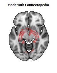
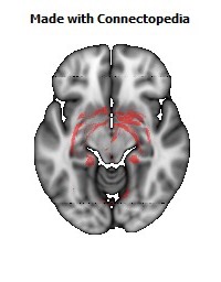
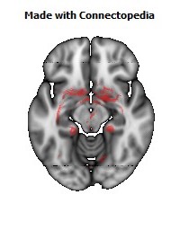
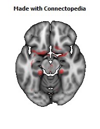
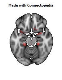
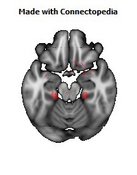

The limbic system (or paleomammalian brain) is a complex set of brain structures that lies on both sides of the thalamus, right under the cerebrum. It is not a separate system, but a collection of structures from the telencephalon, diencephalon, and mesencephalon. It includes the olfactory bulbs, hippocampus, amygdala, anterior thalamic nuclei, fornix, columns of fornix, mammillary body, septum pellucidum, habenular commissure, cingulate gyrus, Parahippocampal gyrus, limbic cortex, and limbic midbrain areas.
The limbic system supports a variety of functions, including emotion, behavior, motivation, long-term memory, and olfaction. It appears to be primarily responsible for emotional life, and it has a great deal to do with the formation of memories.
Some neuroscientists, including Joseph LeDoux, have suggested that the concept of a functionally unified limbic system should be abandoned as obsolete because it is grounded mainly in historical concepts of brain anatomy that are no longer accepted as accurate.
Structure
The limbic system is the set of brain structures that forms the inner border of the cortex. The components of the limbic system located in the cerebral cortex generally have fewer layers than the classical 6-layered neocortex, and are usually classified as allocortex or archicortex.
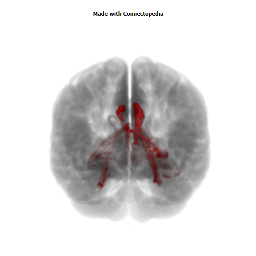
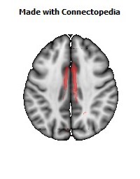
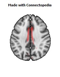
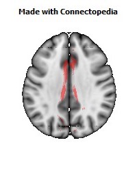
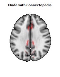
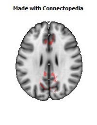
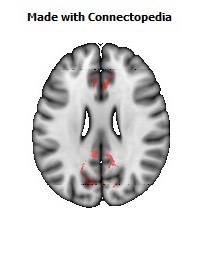
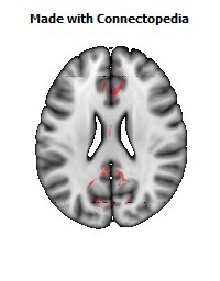
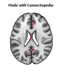
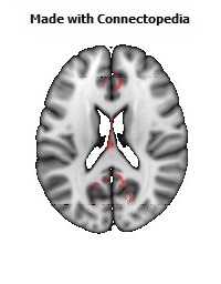
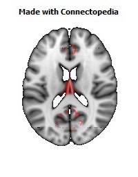
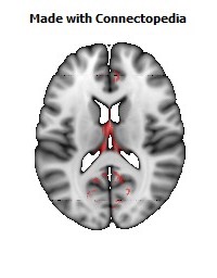
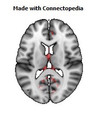
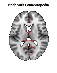
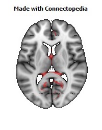
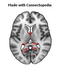
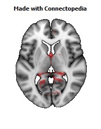
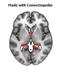
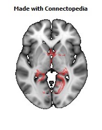
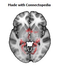
The limbic system is made up of the limbic lobe and other deep-lying structures. The limbic lobe consists of the parahippocampal, cingulate, and subcallosal gyrus, hippocampus, and dentate gyrus. The deep-lying structures include, the hippocampus, amygdala, mammillary body, habenula, anterior thalamic nuclei, and olfactory bulb. The limbic system has four major components that form two C-shaped structures. The first C-shape is made up of the hippocampus and the fornix, while the cingulate and parahippocampal gyri form the second C-shape.
The limbic system includes many structures in the cerebral pre-cortex and sub-cortex of the brain. The term has been used within psychiatry and neurology, although its exact role and definition have been revised considerably since the term was introduced. The following structures are, or have been considered to be, part of the limbic system:
Hippocampus and associated structures:
• Hippocampus: Required for the formation of long-term memories and implicated in maintenance of cognitive maps for navigation. The hippocampus consists of two “horns” that curve back from the amygdalae. It appears to be very important in converting things that are “on one's mind” at the moment (in short-term memory) into things that one will remember for the long run (long-term memory). If the hippocampus is damaged, a person cannot build new memories and lives instead in a strange world where everything he or she experiences just fades away, even while older memories from the time before the damage are untouched (a condition depicted in the films Memento and 50 First Dates).
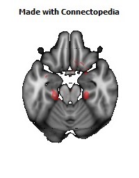
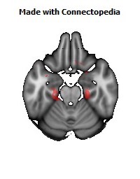
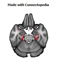
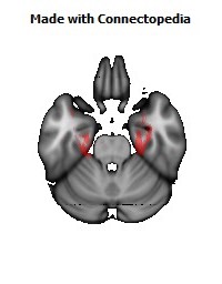
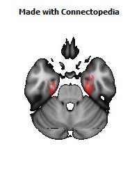
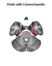
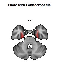
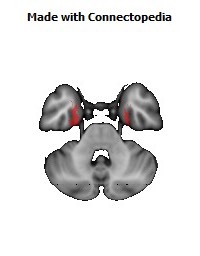
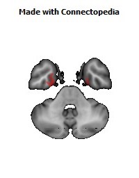
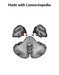
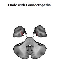
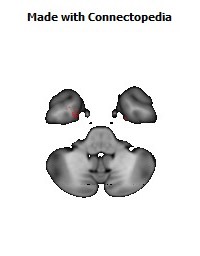
• Amygdala: Involved in signaling the cortex of motivationally significant stimuli such as those related to reward and fear in addition to social functions such as mating. Furthermore, the anatomy of amygdalae are two almond-shaped masses of neurons on either side of the thalamus at the lower end of the hippocampus. The amygdalae stimulate the hippocampus to remember many details surrounding the situation, as well.
• Fornix: is a C-shape bundle of axon that carries signals from the hippocampus to the mammillary bodies and septal nuclei.
• Mammillary body: locates at the ends of the anterior arches of the fornix. It is involved with the process of recognition memory.
• Septal nuclei: Located anterior to the interventricular septum, the septal nuclei provide critical interconnections. The septal area isn't related to the sense of smell, but is the pleasure zone in animals.
Limbic lobe
• Parahippocampal gyrus: Plays a role in the formation of spatial memory.
• Cingulate gyrus: Autonomic functions regulating heart rate, blood pressure and cognitive and attentional processing.
• Dentate gyrus: thought to contribute to new memories.
In addition, these structures are sometimes also considered to be part of the limbic system:
• Entorhinal cortex: Important memory and associative components.
• Piriform cortex: The function of which relates to the olfactory system.
• Fornicate gyrus: Region encompassing the cingulate, hippocampus, and parahippocampal gyrus.
• Nucleus accumbens: Involved in reward, pleasure, and addiction.
• Orbitofrontal cortex: Required for decision making.
Function
The hypothalamus is a part of the limbic system, which is a group of forebrain structures that has the hypothalamus, the amygdala, and the hippocampus. These are involved in motivation, emotion, learning, and memory. The limbic system is where the subcortical structures meet the cerebral cortex. The limbic system operates by influencing the endocrine system and the autonomic nervous system. It is highly interconnected with the nucleus accumbens, the brain's pleasure center, which plays a role in sexual arousal and the "high" derived from certain recreational drugs. These responses are heavily modulated by dopaminergic projections from the limbic system. In 1954, Olds and Milner found that rats with metal electrodes implanted into their nucleus accumbens, as well as their septal nuclei, repeatedly pressed a lever activating this region, and did so in preference to eating and drinking, eventually dying of exhaustion. The limbic system also includes the basal ganglia. The basal ganglia are a set of subcortical structures that directs intentional movements. The basal ganglia are located near the thalamus and hypothalamus. They receive input from the cerebral cortex, which sends outputs to the motor centers in the brain stem. A part of the basal ganglia called the striatum controls posture and movement. Recent studies indicate that, if there is an inadequate supply of dopamine, the striatum is affected, which can lead to visible behavioral symptoms of Parkinson’s. The limbic system is also tightly connected to the prefrontal cortex. Some scientists contend that this connection is related to the pleasure obtained from solving problems. To cure severe emotional disorders, this connection was sometimes surgically severed, a procedure of psychosurgery, called a prefrontal lobotomy (this is actually a misnomer). Patients having undergone this procedure often became passive and lacked all motivation. The limbic system is often classified as a “cerebral structure”. This structure is closely linked to olfaction, emotions, drives, autonomic regulation, memory, and pathologically to encephalopathy, epilepsy, psychotic symptoms, cognitive defects. The functional relevance of the limbic system has proven to serve many different functions such as affects/emotions, memory, sensory processing, time perception, attention, consciousness, instincts, autonomic/vegetative control, and actions/motor behavior. Some of the disorders associated with the limbic system are epilepsy and schizophrenia.
Hippocampus
Spatial memory
The hippocampus has been demonstrated to be involved in various processes of cognition. The first and most widely researched area concerns memory, spatial memory in particular. Spatial memory was found to have many sub-regions in the hippocampus, such as the dentate gyrus (DG) in the dorsal hippocampus, the left hippocampus, and the parahippocampal region. The dorsal hippocampus was found to be an important component for the generation of new neurons, called adult-born granules (GC), in adolescence and adulthood. These new neurons contribute to pattern separation in spatial memory, increasing the firing in cell networks, and overall causing stronger memory formations. While the dorsal hippocampus is involved in spatial memory formation, the left hippocampus is a participant in the recall of these spatial memories. Eichenbaum and his team found, when studying the hippocampal lesions in rats, that the left hippocampus is “critical for effectively combining the ‘what, ‘when,’ and ‘where’ qualities of each experience to compose the retrieved memory.” This makes the left hippocampus a key component in the retrieval of spatial memory. However, Spreng found that the left hippocampus is, in fact, a general concentrated region for binding together bits and pieces of memory composed not only by the hippocampus, but also by other areas of the brain to be recalled at a later time. Eichenbaum’s research in 2007 also demonstrates that the parahippocampal area of the hippocampus is another specialized region for the retrieval of memories just like the left hippocampus.
Learning
The hippocampus, over the decades, has also been found to have a huge impact in learning. CurlikShors examined the effects of neurogenesis in the hippocampus and its effects on learning. This researcher and his team employed many different types of mental and physical training on their subjects, and found that the hippocampus is highly responsive to these latter tasks. Thus, they discovered an upsurge of new neurons and neural circuits in the hippocampus as a result of the training, causing an overall improvement in the learning of the task. This neurogenesis contributes to the creation of adult-born granules cells (GC), cells also described by Eichenbaum in his own research on neurogenesis and its contributions to learning. The creation of these cells exhibited “enhanced excitability” in the dentate gyrus (DG) of the dorsal hippocampus, impacting the hippocampus and its contribution to the learning process.
Hippocampus damage
Damage relayed to the hippocampal region of the brain has reported vast effects on overall cognitive functioning, particularly memory such as spatial memory. As previously mentioned, spatial memory is a cognitive function greatly intertwined with the hippocampus. While damage to the hippocampus may be a result of a brain injury or other injuries of that sort, researchers particularly investigated the effects that high emotional arousal and certain types of drugs had on the recall ability in this specific memory type. In particular, in a study performed by Parkard, rats were given the task of correctly making their way through a maze. In the first condition, rats were stressed by shock or restraint which caused a high emotional arousal. When completing the maze task, these rats had an impaired effect on their hippocampal-dependent memory when compared to the control group,. Then, in a second condition, a group of rats were injected with anxiogenic drugs. Like the former these results reported similar outcomes, in that hippocampal-memory was also impaired. Studies such as these reinforce the impact that the hippocampus has on memory processing, in particular the recall function of spatial memory. Furthermore, impairment to the hippocampus can occur from prolonged exposure to stress hormones such as Glucocorticoids (GCs), which target the hippocampus and cause disruption in explicit memory.
In an attempt to curtail life threatening epileptic seizures, 27-year-old Henry Gustave Molaison underwent bilateral removal of almost all of his hippocampus in 1953. Over the course of fifty years he participated in thousands of tests and research projects that provided specific information on exactly what he had lost. Semantic and episodic events faded within minutes, having never reached his long term memory, yet emotions, unconnected from the details of causation, were often retained. Dr. Suzanne Corkin who worked with him for 46 years until his death described the contribution of this tragic "experiment" in her 2013 book.
Amygdala
Episodic-autobiographical memory (EAM) networks
The amygdala, another integrative part of the limbic system, is also involved in many cognitive processes. Just as in the hippocampus, memory seems to be impacted by processes in the amygdale; however, it is not spatial memory as in the hippocampus, but episodic-autobiographical memory (EAM) networks. The amygdala, as researched by Markowitsch, was found to be responsible for the encoding, storage, and retrieval of these types of memories. To delve deeper into these types of processes by the amygdala, Markowitsch and his team provided extensive evidence through investigations that the “amygdala’s main function is to charge cues so that mnemonic events of a specific emotional significance can be successfully searched within the appropriate neural nets and re-activated.” These cues for emotional events created by the amygdala encompass the EAM networks previously mentioned.
Attentional and emotional processes
Besides memory, the amygdala also seems to be an important brain region involved in attentional and emotional processes. First, to define attention in cognitive terms, attention is the ability to hone in on some stimuli while ignoring others. Thus, the amygdala seems to be an important structure in this ability. Foremost, however, this structure was historically thought to be linked to fear, allowing the individual to take action to rid that fear in some sort. However, as time has gone by, researchers such as Pessoa, generalized this concept with help from evidence of EEG recordings, and concluded that the amygdala helps an organism to define a stimulus and therefore respond accordingly. However, when the amygdala was initially thought to be linked to fear, this gave way for research in the amygdala for emotional processes. Kheirbek demonstrated research that the amygdala is involved in emotional processes, in particular the ventral hippocampus. He described the ventral hippocampus as having a role in neurogenesis and the creation of adult-born granule cells (GC). These cells not only were a crucial part of neurogenesis and the strengthening of spatial memory and learning in the hippocampus but also appear to be an essential component in the amygdala. A deficit of these cells, as Pessoa (2009) predicted in his studies, would result in low emotional functioning, leading to high retention rate of mental diseases, such as anxiety disorders.
Social processing
Social processing is an area of cognition specific to the amygdala. To be specific, the evaluation of faces in social processing is of particular importance. In a study done by Todorov, fMRI tasks were performed with participants to evaluate whether the amygdala was involved in the general evaluation of faces. After the study, Todorov concluded from his fMRI results that the amygdala did indeed play a key role in the general evaluation of faces. However, in a study performed by researchers Koscik and his team, the trait of truthworthiness was particularly examined in the evaluation of faces. They investigated how brain damage to the amygdala played a role in truthworthiness, and found that individuals that suffered damage tended to confuse trust and betrayal, and thus placed trust in those having done them wrong. So Koscik demonstrated that the amygdala was involved in evaluating the truthworthiness of an individual. Yet, a man named Rule, along with his colleagues, expanded on the idea of the amygdala in its critique of truthworthiness in others and performed a study in 2009 in which he examined the amygdala in its role of evaluating general first impressions and relating them to real-world outcomes with his study involving first impressions of CEOs. Rule demonstrated that while the amygdala did play a role in the evaluation of truthworthiness, as observed by Koscik in his own research two years later in 2011, the amygdala played a generalized role in the overall evaluation of first impression of faces. This latter conclusion, along with Todorov’s study on the amygdala’s role in general evaluations of faces and Koscik’s research on truthworthiness and the amygdala, further solidified evidence that the amygdala plays a role in overall social processing.
Evolution
Paul D. MacLean, as part of his triune brain theory, hypothesized that the limbic system is older than other parts of the fore-brain, and that it developed to manage fight or flight circuitry, which is an evolutionary necessity for reptiles as well as humans. MacLean postulated that the human brain has evolved three components, that evolved successively, with more recent components developing at the top/front. These components are, respectively:
1 - The archipallium or primitive ("reptilian") brain, comprising the structures of the brain stem - medulla, pons, cerebellum, mesencephalon, the oldest basal nuclei - the globus pallidus and the olfactory bulbs.
2 - The paleopallium or intermediate ("old mammalian") brain, comprising the structures of the limbic system.
3 - The neopallium, also known as the superior or rational ("new mammalian") brain, comprises almost the whole of the hemispheres (made up of a more recent type of cortex, called neocortex) and some subcortical neuronal groups. It corresponds to the brain of the superior mammals, thus including the primates and, as a consequence, the human species.
According to Maclean, each of the components, although connected with the others, retained "their peculiar types of intelligence, subjectivity, sense of time and space, memory, mobility and other less specific functions".
However, while the categorizaton into structures is reasonable, the recent studies of the limbic system of tetrapods, both living and extinct, have challenged several aspects of this hypothesis, notably the accuracy of the terms "reptilian" and "old mammalian". The common ancestors of reptiles and mammals had a well-developed limbic system in which the basic subdivisions and connections of the amygdalar nuclei were established. further, birds, which evolved from the dinosaurs, which in turn evolved separately but around the same time as the mammals, have a well-developed limbic system. While the anatomic structures of the limbic system are different in birds than in mammals, there are functional equivalents.
Clinical significance
Damage to the structures of limbic system results in conditions like Alzheimer's disease, anterograde amnesia, retrograde amnesia, and Kluver-Bucy syndrome.
History
The limbic system is a term that was introduced in 1952 by physician and neuroscientist, Paul D. MacLean.
The first evidence that the limbic system was responsible for the cortical representation of emotions was discovered in 1939, by Paul Kluver and Heinrich Bucy. Kluver and Bucy, after much research, demonstrated that the bilateral removal of the temporal lobes in monkeys created an extreme behavioral syndrome. After performing a temporal lobectomy, the monkeys showed a decrease in aggression. The animals revealed a reduced threshold to visual stimuli, and were thus unable to recognize objects that were once familiar. The French physician Paul Broca first called this part of the brain le grand lobe limbique in 1878. He examined the differentiation between deeply recessed cortical tissue and underlying, subcortical nuclei. However, most of its putative role in emotion was developed only in 1937 when the American physician James Papez described his anatomical model of emotion, the Papez circuit. Paul D. MacLean expanded these ideas to include additional structures in a more dispersed "limbic system," more on the lines of the system described above. The term was formally introduced by Paul D. MacLean in 1952. Paul D. MacLean developed the intriguing theory of the “triune brain” to explain its evolution and to try to reconcile rational human behavior with its more primal and violent side. He became interested in the brain’s control of emotion and behavior. After initial studies of brain activity in epileptic patients, he turned to cats, monkeys, and other models, using electrodes to stimulate different parts of the brain in conscious animals. Furthermore, He then recorded the animals’ responses and, in the 1950s, he began to trace individual behaviors like aggression and sexual arousal to their physiological sources. He analyzed the brain’s center of emotions, the limbic system, and described an area that includes structures called the hippocampus and amygdala. Developing observations made by Dr. James W. Papez, he determined that the limbic system had evolved in early mammals to control fight-or-flight responses and react to both emotionally pleasurable and painful sensations. The concept is now broadly accepted in neuroscience. Additionally, Dr. MacLean said that the idea of the limbic system leads to a recognition that its presence “represents the history of the evolution of mammals and their distinctive family way of life.”In the 1960s, Dr. MacLean enlarged his theory to address the human brain’s overall structure and divided its evolution into three parts, an idea that he termed the triune brain. In addition to identifying the limbic system, he pointed to a more primitive brain called the R-complex, related to reptiles, which controls basic functions like muscle movement and breathing. The third part, the neocortex, controls speech and reasoning and is the most recent evolutionary arrival. The concept of the limbic system has since been further expanded and developed by Walle Nauta, Lennart Heimer and others.
Etymology
The term "limbic" comes from the Latin limbus, for "border" or "edge", or, particularly in medical terminology, a border of an anatomical component. Paul Broca coined the term based on its physical location in the brain, sandwiched between two functionally different components.
Academic dispute
There is recent controversy over the use of the term limbic system, with scientists such as LeDoux arguing that the term be considered obsolete and abandoned. Originally, the limbic system was believed to be the emotional center of the brain, with cognition being the business of the neocortex. However, cognition depends on acquisition and retention of memories, in which the hippocampus, a primary limbic structure, is involved: hippocampus damage causes severe cognitive (memory) deficits. More important, the "boundaries" of the limbic system have been repeatedly redefined because of advances in neuroscience. Therefore, while it is true that limbic structures are more closely related to emotion, one must think of the brain as an integrated whole.
Neuroanatomy of memory
Subcortical structures
Hippocampus
The hippocampus is a structure in the brain that has been associated with various memory functions. It is part of the limbic system, and lies next to the medial temporal lobe. It is made up of two structures, the Ammon’s Horn, and the Dentate gyrus, each containing different types of cells.
Cognitive maps
There is evidence that the hippocampus contains cognitive maps in humans. In one study, single-cell recordings were taken from electrodes implanted in a rat’s hippocampus, and it was found that certain neurons responded strongly only when the rat was in certain locations. These cells are called place cells, and collections of these cells can be considered to be mental maps. Individual place cells do not only respond to one unique area only however, the patterns of activation of these cells overlap to form layered mental maps within the hippocampus. A good analogy is the example of the same television or computer screen pixels being used to light up any trillions of possible combinations to produce images, just as the place cells can be used in any multiple possible combinations to represent mental maps. The hippocampus’ right side is more oriented towards responding to spatial aspects, whereas the left side is associated with other context information. Also, there is evidence that experience in building extensive mental maps, such as driving a city taxi for a long time (since this requires considerable memorization of routes), can increase the volume of one’s hippocampus.
Encoding
Damage to the hippocampus and surrounding area can cause anterograde amnesia, the inability to form new memories. This implies that the hippocampus is important not only for storing cognitive maps, but for encoding memories.
The hippocampus is also involved in memory consolidation, the slow process by which memories are converted from short to long term memory. This is supported by studies in which lesions are applied to rat hippocampi at different times after learning. The process of consolidation may take up to a couple years.
It has also been found that it is possible to form new semantic memories without the hippocampus, but not episodic memories, which means that explicit descriptions of actual events (episodic) cannot be learned, but some meaning and knowledge is gained from experiences (semantic).
Cerebellum
The cerebellum ("little brain") is a structure located at the rear of the brain, near the spinal cord. It looks like a miniature version of the cerebral cortex, in that it has a wavy, or convoluted surface.
Unlike the hippocampus which is involved in the encoding of complex memories, the cerebellum plays a role in the learning of procedural memory, and motor learning, such as skills requiring co-ordination and fine motor control. An example of a skill requiring procedural memory would be playing a musical instrument, or driving a car or riding a bike. Individuals with transient global amnesia that have difficulty forming new memories and/or remembering old events may sometimes retain the ability to perform complex musical pieces, suggesting that procedural memory is completely dissociated from conscious memory, also known as explicit memory.
This separation makes sense if the cerebellum, which is far removed from the hippocampus, is responsible for procedural learning. The cerebellum is more generally involved in motor learning, and damage to it can result in problems with movement, specifically it is considered to co-ordinate timing and accuracy of movements, and to make long-term changes (learning) to improve these skills.
Amygdala
Located below the hippocampus in the medial temporal lobes are two amygdalae (singular "amygdala"). The amygdalae are associated with both emotional learning and memory, as it responds strongly to emotional stimuli, especially fear. These neurons assist in encoding emotional memories and enhancing them. This process results in emotional events being more deeply and accurately encoded into memory. Lesions to the amygdalae in monkeys have been shown to impair motivation, as well as the processing of emotions.
Memory of fear conditioning
Pavlovian conditioning tests have shown the active role of the amygdala in fear conditioning in rats. Research involving lesions to the basolateral nucleus have shown a strong association with memories involving fear. The central nucleus is linked with the behavioral responses that are dependent on the basolateral’s reaction to fear. The central nucleus of the amygdala is also linked to emotions and behaviors motivated by food and sex.
Memory consolidation
Emotional experiences and events are somewhat fragile and take a while to be completely set into memory. This slow process, referred to as consolidation, allows emotions to influence the way the memory is stored.
The amygdala is involved in memory consolidation, which is the process of transferring information that is currently in working memory into ones long-term memory. This process is also known as memory modulation. The amygdala works to encode recent emotional information into memory. Memory research has shown that the greater ones emotional arousal level at the time of the event, the greater the chance that the event will be remembered. This may be due to the amygdala enhancing the emotional aspect of the information during encoding, causing the memory to be processed at a deeper level and therefore, more likely to withstand forgetting.
Basal ganglia and motor memory
The basal ganglia are a group of nuclei which are located in the medial temporal lobe, above the thalamus and connected to the cerebral cortex. Specifically, the basal ganglia includes the subthalamic nucleus, substantia nigra, the globus pallidus, the ventral striatum and the dorsal striatum, which consists of the putamen and the caudate nucleus. The basic functions of these nuclei deal with cognition, learning, and motor control and activities. The basal ganglia are also associated with learning, memory, and unconscious memory processes, such as motor skills and implicit memory. Particularly, one division within the ventral striatum, the nucleus accumbens core, is involved in the consolidation, retrieval and reconsolidation of drug memory.
The caudate nucleus is thought to assist in learning and memory of associations taught during operant conditioning. Specifically, research has shown that this part of the basal ganglia plays a role in acquiring stimulus-response habits, as well as in solving sequence tasks.
Damage to the basal ganglia has been linked to dysfunctional learning of motor and perceptual-motor skills. Most disorders that are associated with damage to these areas of the brain involve some type of motor dysfunction, as well as trouble with mental switching between tasks in working memory. Such symptoms are often present in those who suffer from dystonia, athymhormic syndrome, Fahr's syndrome, Huntington's disease or Parkinson's disease. Huntington's and Parkinson's disease involve both motor deficits and cognitive impairment.
Cortical structures
Frontal lobe
The frontal lobes are located at the front of each cerebral hemisphere and positioned anterior to the parietal lobes. It is separated from the parietal lobe by the primary motor cortex, which controls voluntary movements of specific body parts associated with the precentral gyrus. The cortex here serves our ability to plan the day, organize work, type a letter, pay attention to details and control the movements of your arms and legs. It also contributed to your personality and behaviour.
When considering the frontal lobes in regards to memory, we see that it is very important in the coordination of information. Therefore, the frontal lobes are important in working memory. For example, when you are thinking about how to get to a mall you have never been to before, you combine various bits of knowledge you already have: the layout of the city the mall is in, information from a map, knowledge of traffic patterns in that area and conversations with your friends about the location of the mall. By actively using all of this information, you can determine the best route for you to take. This action involves the controlled use of information in working memory, coordinated by the frontal lobes.
The frontal lobes help a person select out memories that are most relevant on a given occasion. It can coordinate various types of information into a coherent memory trace. For example, the knowledge of the information itself, as well as knowing where information came from must be put together into a single memory representation; this is called source monitoring. Sometimes we experience situations where information becomes separated, such as when we recall something, but cannot remember where we remember it from; this is referred to as a source monitoring error.
The frontal lobes are also involved in the ability to remember what we need to do in the future; this is called prospective memory.
Temporal lobe
The temporal lobes are a region of the cerebral cortex that is located beneath the Sylvian fissure on both the left and right hemispheres of the brain. Lobes in this cortex are more closely associated with memory and in particular autobiographical memory.
The temporal lobes are also concerned with recognition memory. This is the capacity to identify an item as one that was recently encountered. Recognition memory is widely viewed as consisting of two components, a familiarity component (i.e. Do I know this person waving at me?) and a recollective component (i.e. That is my friend Julia, from evolutionary psychology class).
Damage to the temporal lobe can affect an individual in a litany of ways ranging from: disturbance of auditory sensation and perception, disturbance of selective attention of auditory and visual input, disorders of visual perception, impaired organization and categorization of verbal material, disturbance of language comprehension, and altered personality.
In regard to memory, temporal lobe damage can impair long-term memory. Thus, general semantic knowledge or more personal episodic memories of one’s childhood could be affected.
Parietal lobe
The parietal lobe is located directly behind the central sulcus, superior to the occipital lobe and posterior to the frontal lobe, visually at the top of the back of the head. The make up of the parietal lobe is defined by four anatomical boundaries in the brain, providing a division of all the four lobes.
The parietal lobe has many functions and duties in the brain and its main functioning can be divided down into two main areas: (1) sensation and perception (2) constructing a spatial coordinate system to represent the world around us. The parietal lobe helps us to mediate attention when necessary and provides spatial awareness and navigational skills. Also, it integrates all of our sensory information (touch, sight, pain etc.) to form a single perception. Parietal lobe gives the ability to focus our attention on different stimuli at the same time, PET scans show high activity in the parietal lobe when participates being studied were asked to focus their attention at two separate areas of attention. Parietal lobe also assists with verbal short term memory and damage to the supramarginal gyrus cause short term memory loss.
Damage to the parietal lobe results in the syndrome ‘neglect’ which is when patients treat part of their body or objects in their visual field as though it never existed. Damage to the left side of the parietal lobe can result in what is called Gerstmann syndrome. It includes right-left confusion, difficulty with writing (agraphia) and difficulty with mathematics (acalculia). It can also produce disorders of language (aphasia) and the inability to perceive objects. Damage to the right parietal lobe can result in neglecting part of the body or space (contralateral neglect), which can impair many self-care skills such as dressing and washing. Right side damage can also cause difficulty in making things (constructional apraxia), denial of deficits (anosognosia) and drawing ability. Neglect syndrome tends to be more prevalent on the right side of the parietal lobe, because the right mediates attention to both the left and right fields. Damage in the somatic sensory cortex results in loss of perception of bodily sensations, namely sense of touch.
Occipital lobe
The occipital lobe is the smallest of all four lobes in the human cerebral cortex and located in the rearmost part of the skull and considered to be part of the forebrain. The occipital lobe sits directly above the cerebellum and is situated posterior to the Parieto-occipital sulcus, or parieto-occipital sulcus. This lobe is known as the centre of the visual perception system, the main function of the occipital lobe is that of vision.
Retinal sensors send signals through the optic tract to the Lateral geniculate nucleus. Once the Lateral Geniculate Nucleus receives the information it is sent down the primary visual cortex where it is organized and sent down one of two possible path ways; dorsal or ventral stream. The ventral stream is responsible for object representation and recognition and is also commonly known as the "what" stream. The dorsal stream is responsible for guiding our actions and recognizing where objects are in space, commonly known as the "where" or "how" stream. Once in the information is organized and sent through the pathways it continues to the other areas of the brain responsible for visual processing.
The most important function of the Occipital lobe is vision. Due to the positioning of this lobe at the back of the head it is not susceptible to much injury but any significant damage to the brain can cause a variety of damage to our visual perception system. Common problems in the occipital lobe are field defects and scotomas, movement and colour discrimination, hallucinations, illusions, inability to recognize words and inability to recognize movement. A study was done in which patients suffered from a tumour on the occipital lobe and the results shows that the most frequent consequence was contralateral damage to the visual field. When damage occurs in the occipital lobe it is most common to see the effects on the opposite side of the brain. Since the brain regions are so specialized in their functioning damages done to specific areas of the brain can cause specific type of damage. Damage to the left side of the brain can lead to language discrepancies, i.e. difficulty in properly identifying letters, numbers and words, inability to incorporate visual stimuli to comprehend multiple ways an object can be found. Right side damage causes non-verbal problems, i.e. identifying geometric shapes, perception of figures and faces. In almost all regions of the brain left side damage leads to general language problems whereas right side damage leads to general perception and problem solving skills.
Damage to the cortex
Many studies of different disease and disorders that have symptoms of memory loss have provided reinforcing evidence to the study of the anatomy of the brain and which parts are more utilized in memory.
Frontotemporal lobar degeneration and memory
Frontotemporal lobar degeneration (FTLD) is a common form of dementia due to the degeneration of the frontal and temporal lobes. Studies have found significant decreases in the essential needs for proper functioning in these lobes. The autobiographical domain in memory is largely affected by this disease. In one study, FTLD patients were interviewed and asked to describe a significant event from five different periods of their lives. Using the interview and different methods of imaging, the experimenters hoped to find links between patterns of brain volume loss and performance in the interview.
Through image processing, patterns of significant reduced parenchymal volumes that encompass the frontal and temporal lobes were found. Through comparison to a control group of patients it was found that parenchymal volumes increased during episodic recall, and decreased during semantic recall. The experimenters discussed that lifespan autobiographical episodic recall was largely damaged in FTLD patients and semantic autobiographical memory seemed to be spared.
Parkinson's disease and memory
Parkinson's disease involves both damage to the basal ganglia and certain memory dysfunctions, suggesting that the basal ganglia are involved in specific types of memory. Those who have this disease have problems with both their working memory and spatial memory.
Most people can instantly and easily use visual-spatial memory to remember locations and pictures, but a person with Parkinson's disease would find this difficult. He or she would also have trouble encoding this visual and spatial information into long-term memory. This suggests that the basal ganglia work in both encoding and recalling spatial information.
People with Parkinson's disease display working memory impairment during sequence tasks and tasks involving events in time. They also have difficulty in knowing how to use their memory, such as when to change strategies or maintain a train of thought.