
The medial lemniscus, also known as Reil's band or Reil's ribbon, is a pathway in the brainstem that carries sensory information from the gracile and cuneate nuclei to the thalamus.
Path
After neurons carrying proprioceptive or touch information synapse at the gracile and cuneate nuclei, axons from secondary neurons decussate at the level of the medulla and travel up the brainstem as the medial lemniscus on the contralateral (opposite) side. It is part of the posterior column-medial lemniscus system, which transmits touch, vibration sense, as well as the pathway for proprioception.
The medial lemniscus carries axons from most of the body and synapses in the ventral posterolateral nucleus of the thalamus, at the level of the mamillary bodies. Sensory axons transmitting information from the head and neck via the trigeminal nerve synapse at the ventral posteromedial nucleus of the thalamus.
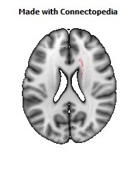
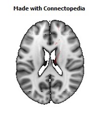
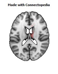
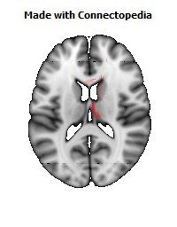
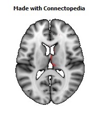
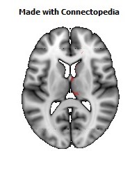
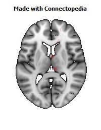
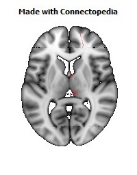
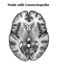
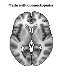
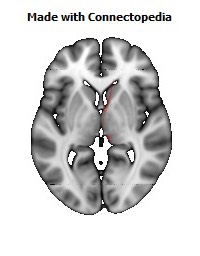
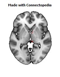
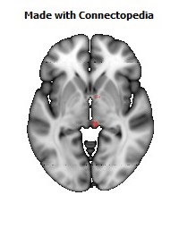
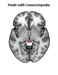
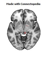
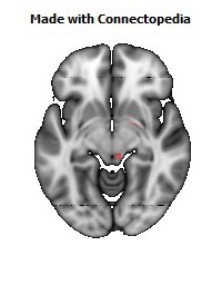
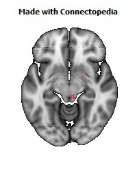
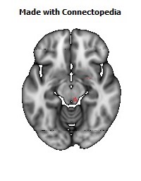
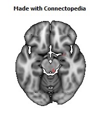
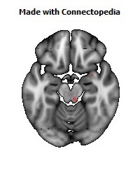
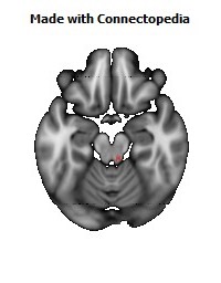
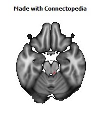
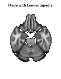
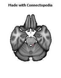
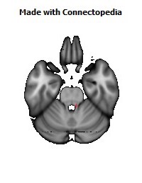
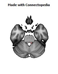
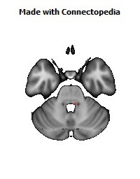
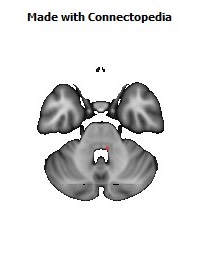
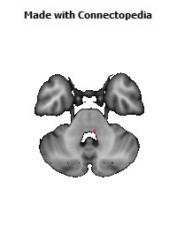
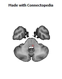
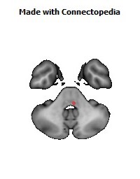
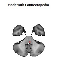
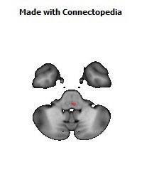
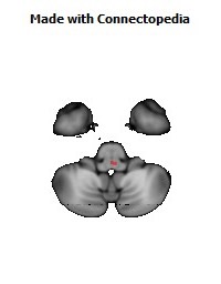
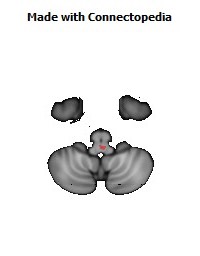
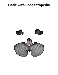
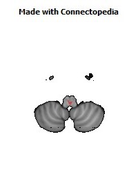
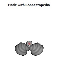
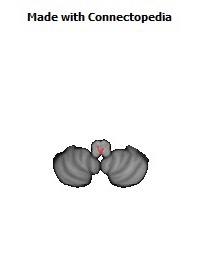
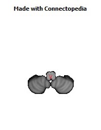
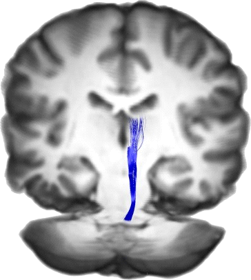
Location of the medial lemniscus through the brainstem
• The cuneate and gracile nuclei reside at the closed (lower) medulla, so the lemniscus isn't formed at this level. Fibres from these nuclei will pass to the contralateral side of the brainstem, as the internal arcuate fibres.
• At the open medulla (further up the brainstem), the medial lemniscus contains axons from the trigeminal nerve (which supplies the head region), as well as the arms and legs. It sits very close to the midline, at the same orientation of the midline, with head fibres more dorsal (closer to the back), towards the fourth ventricle.
• By mid-pons, the medial lemniscus has rotated. Fibres from the head are medial, fibres from the leg are lateral.
The orientation in the midbrain is similar to that in the pons.
The posterior column-medial lemniscus pathway, PCML for short, (also known as the dorsal column, dorsal column-medial lemniscus pathway or the dorsal white column-medial lemniscus system) is the sensory pathway responsible for transmitting fine touch, vibration and conscious proprioceptive information from the body to the cerebral cortex;[1] as well as tactile pressure, barognosis, graphesthesia, stereognosis, recognition of texture, kinesthesia and two-point discrimination.
The name comes from the two structures that the sensation travels up: the posterior (or dorsal) columns of the spinal cord, and the medial lemniscus in the brainstem. Because the posterior columns are also called dorsal columns, the pathway is often called the dorsal column-medial lemniscus system, or DCML for short.
The PCML pathway is composed of rapidly conducting, large, myelinated fibers.
Journey
Discriminative sensation is well developed in the fingers of humans, and allows us to feel fine textures and determine what an unknown object in our hands is without looking at it (stereognosis).
First neuron
This fine sensation is detected by Meissner's corpuscles that lie in the dermis of the skin close to the epidermis. When these structures are stimulated by slight pressure, an action potential is started. Alternatively, proprioceptive muscle spindles and other skin surface touch receptors such as Merkel cells, Ruffini endings, Pacinian corpuscles, and hair follicle receptors (peritrichial endings) may involve the first neuron in this pathway.
The action potential travels up an axon (the cell body of the neuron will be in a dorsal root ganglion). (The neurons are classified as pseudo-unipolar, so they are regarded as having just one long process, which includes both a peripheral branch dendrite and central branch axon.) So the sensation travels from the skin, along the axon, past the neuronal cell body, and into the dorsal column of the spinal cord.
Tracts of the spinal cord, showing the involved tracts of the Dorsal Column Medial Lemniscus System labeled at right.
The axons continue inside the spinal cord, running up the posterior (dorsal) column. Axons from the lower body are most medial (closer to the midline), and run in the gracile tract of the spinal column. Sensory axons from the upper body enter the spinal cord later, specifically from T6 on up, so are more lateral and travel up the cuneate tract.
At the level of the closed medulla oblongata, these axons synapse with neurons in the gracile nucleus and cuneate nucleus.
Second neuron
The secondary neurons (that start in the nuclei) cross over to the other side of the medulla (as internal arcuate fibres) to form the medial lemniscus. This crossing over is commonly referred to as the sensory decussation.
At the medulla, the medial lemniscus is orientated perpendicular to the way the fibres travelled in the posterior columns. For example, in the columns, lower limb is medial, upper limb is more lateral. At the medial lemniscus, axons from the leg are more ventral, and axons from the arm are more dorsal. Fibres from the trigeminal nerve (supplying the head) come in dorsal to the arm fibres, and travel up the lemniscus too.
The medial lemniscus rotates 90 degrees at the pons. The secondary axons from neurons giving sensation to the head, stay at around the same place, while the leg axons move outwards.
The axons travel up the rest of the brainstem, and synapse at the thalamus (at the ventral posterolateral nucleus for sensation from the neck, trunk, and extremities, and at the ventral posteromedial nucleus for sensation from the head).
Third neuron
Neurons starting in the thalamus travel up the posterior limb of the internal capsule, and again head and leg swap relative positions. The axons synapse in the primary sensory cortex, with lower body sensation most medial (e.g., the paracentral lobule) and upper body more lateral.
Test
The pathway is tested with the Romberg's test. Lesions to the posterior column-medial lemniscus pathway below the decussation of its fibers produce loss of sensation on the same side of the body as the lesion. Above the decussation produces loss of sensation on the opposite side of the body than the lesion.